 Movie
Movie Controller

[English] 日本語
 Yorodumi
- PDB-7jh0: Crystallographic structure of glyceraldehyde-3-phosphate dehydrog... -
+ Open data

- Basic information
| Entry | Database: PDB / ID: 7jh0 | ||||||
|---|---|---|---|---|---|---|---|
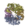
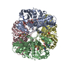
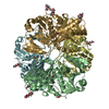
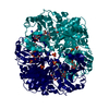
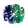
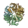
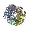
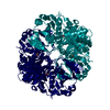
| Title | Crystallographic structure of glyceraldehyde-3-phosphate dehydrogenase from Schistosoma mansoni | ||||||
 Components Components | (Glyceraldehyde-3-phosphate ...) x 2 | ||||||
 Keywords Keywords | OXIDOREDUCTASE | ||||||
| Function / homology |  Function and homology information Function and homology informationglyceraldehyde-3-phosphate dehydrogenase (phosphorylating) / glyceraldehyde-3-phosphate dehydrogenase (NAD+) (phosphorylating) activity / glycolytic process / glucose metabolic process / NAD binding / NADP binding / cytosol Similarity search - Function | ||||||
| Biological species |  | ||||||
| Method |  X-RAY DIFFRACTION / SYNCHROTRON / MOLECULAR REPLACEMENT / Resolution: 2.51 Å X-RAY DIFFRACTION / SYNCHROTRON / MOLECULAR REPLACEMENT / Resolution: 2.51 Å | ||||||
 Authors Authors | Boreiko, S. / Silva, M. / Iulek, J. | ||||||
 Citation Citation | Journal: Biochimie / Year: 2021 Title: Structure determination and analyses of the GAPDH from the parasite Schistosoma mansoni, the first one from a platyhelminth. Authors: Boreiko, S. / Silva, M. / Iulek, J. | ||||||
| History |
|
- Structure visualization
| Structure viewer | Molecule: MolmilJmol/JSmol |
|---|
- Downloads & links
-Download
| PDBx/mmCIF format | 7jh0.cif.gz | 510.9 KB | Display | PDBx/mmCIF format |
|---|---|---|---|---|
| PDB format | pdb7jh0.ent.gz | 422.7 KB | Display | PDB format |
| PDBx/mmJSON format | 7jh0.json.gz | Tree view | PDBx/mmJSON format | |
| Others |  Other downloads Other downloads |
-Validation report
| Arichive directory | https://data.pdbj.org/pub/pdb/validation_reports/jh/7jh0ftp://data.pdbj.org/pub/pdb/validation_reports/jh/7jh0 | HTTPS FTP |
|---|
-Related structure data
| Related structure data |  4k9dS S: Starting model for refinement |
|---|---|
| Similar structure data |


-Links
 PDBj
PDBj
- Assembly
| Deposited unit | 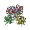
| ||||||||
|---|---|---|---|---|---|---|---|---|---|
| 1 |
| ||||||||
| Unit cell |
|
-Components
-Glyceraldehyde-3-phosphate ... , 2 types, 4 molecules ABCD
| #1: Protein | Mass: 36503.578 Da / Num. of mol.: 3 Source method: isolated from a genetically manipulated source Source: (gene. exp.)  References: UniProt: P20287, glyceraldehyde-3-phosphate dehydrogenase (phosphorylating) #2: Protein | | Mass: 36455.594 Da / Num. of mol.: 1 Source method: isolated from a genetically manipulated source Source: (gene. exp.) References: UniProt: P20287, glyceraldehyde-3-phosphate dehydrogenase (phosphorylating) |
|---|
-Non-polymers , 5 types, 210 molecules 


| #3: Chemical |  Mass: 92.094 Da / Num. of mol.: 2 / Source method: obtained synthetically / Formula: C3H8O3 Mass: 92.094 Da / Num. of mol.: 2 / Source method: obtained synthetically / Formula: C3H8O3#4: Chemical |  Mass: 150.173 Da / Num. of mol.: 2 / Source method: isolated from a natural source / Formula: C6H14O4 Mass: 150.173 Da / Num. of mol.: 2 / Source method: isolated from a natural source / Formula: C6H14O4#5: Chemical |  Mass: 106.120 Da / Num. of mol.: 2 / Source method: isolated from a natural source / Formula: C4H10O3 Mass: 106.120 Da / Num. of mol.: 2 / Source method: isolated from a natural source / Formula: C4H10O3#6: Chemical | ChemComp-EDO / |  Mass: 62.068 Da / Num. of mol.: 1 / Source method: obtained synthetically / Formula: C2H6O2 Mass: 62.068 Da / Num. of mol.: 1 / Source method: obtained synthetically / Formula: C2H6O2#7: Water | ChemComp-HOH / | Mass: 18.015 Da / Num. of mol.: 203 / Source method: isolated from a natural source / Formula: H2O |
|---|
-Details
| Has ligand of interest | N |
|---|---|
| Has protein modification | Y |
-Experimental details
-Experiment
| Experiment | Method: X-RAY DIFFRACTION / Number of used crystals: 1 |
|---|
- Sample preparation
| Crystal | Density Matthews: 2.65 Å3/Da / Density % sol: 53.54 % |
|---|---|
| Crystal grow | Temperature: 291.15 K / Method: vapor diffusion, hanging drop Details: 0.12 M Ethylene glycols (0.3 M diethyleneglycol, 0.3 M triethyleneglycol, 0.3 M tetraethyleneglycol, 0.3 M pentaethyleneglycol) , 0.1 M Buffer system 1 pH 6.5 (0,1 M MES/Imidazole) and 30% ...Details: 0.12 M Ethylene glycols (0.3 M diethyleneglycol, 0.3 M triethyleneglycol, 0.3 M tetraethyleneglycol, 0.3 M pentaethyleneglycol) , 0.1 M Buffer system 1 pH 6.5 (0,1 M MES/Imidazole) and 30% Mix of precipitants (20% V/V Glicerol e 10% w/V PEG 40,000) |
-Data collection
| Diffraction | Mean temperature: 100 K / Serial crystal experiment: N |
|---|---|
| Diffraction source | Source: SYNCHROTRON / Site: SOLEIL  / Beamline: PROXIMA 1 / Wavelength: 0.978 Å / Beamline: PROXIMA 1 / Wavelength: 0.978 Å |
| Detector | Type: DECTRIS EIGER X 16M / Detector: PIXEL / Date: Feb 6, 2020 |
| Radiation | Protocol: SINGLE WAVELENGTH / Monochromatic (M) / Laue (L): M / Scattering type: x-ray |
| Radiation wavelength | Wavelength: 0.978 Å / Relative weight: 1 |
| Reflection twin | Operator: h,-h-k,-l / Fraction: 0.08 |
| Reflection | Resolution: 2.51→57.76 Å / Num. obs: 51375 / % possible obs: 99.9 % / Redundancy: 21.57 % / CC1/2: 0.999 / Rrim(I) all: 0.172 / Net I/σ(I): 14.35 |
| Reflection shell | Resolution: 2.51→2.57 Å / Mean I/σ(I) obs: 1.13 / Num. unique obs: 3515 / CC1/2: 0.501 / Rrim(I) all: 2.654 / % possible all: 99.9 |
- Processing
| Software |
| ||||||||||||||||||||||||||||||||||||||||
|---|---|---|---|---|---|---|---|---|---|---|---|---|---|---|---|---|---|---|---|---|---|---|---|---|---|---|---|---|---|---|---|---|---|---|---|---|---|---|---|---|---|
| Refinement | Method to determine structure: MOLECULAR REPLACEMENT Starting model: 4K9D Resolution: 2.51→57.76 Å / Cross valid method: THROUGHOUT / σ(F): 1.87 / Phase error: 35.86 / Stereochemistry target values: TWIN_LSQ_F
| ||||||||||||||||||||||||||||||||||||||||
| Solvent computation | Shrinkage radii: 0.9 Å / VDW probe radii: 1.11 Å / Solvent model: FLAT BULK SOLVENT MODEL | ||||||||||||||||||||||||||||||||||||||||
| Displacement parameters | Biso max: 173.41 Å2 / Biso mean: 74.0242 Å2 / Biso min: 35.66 Å2 | ||||||||||||||||||||||||||||||||||||||||
| Refinement step | Cycle: final / Resolution: 2.51→57.76 Å
| ||||||||||||||||||||||||||||||||||||||||
| LS refinement shell | Resolution: 2.51→2.57 Å / Rfactor Rfree error: 0 / Total num. of bins used: 14
| ||||||||||||||||||||||||||||||||||||||||
| Refinement TLS params. | Method: refined / Origin x: -8.9134 Å / Origin y: 11.2973 Å / Origin z: -3.4192 Å
| ||||||||||||||||||||||||||||||||||||||||
| Refinement TLS group |
|
