 Movie
Movie Controller

[English] 日本語
 Yorodumi
- PDB-6ynd: GAPDH purified from the supernatant of HEK293F cells: crystal for... -
+ Open data

- Basic information
| Entry | Database: PDB / ID: 6ynd | |||||||||
|---|---|---|---|---|---|---|---|---|---|---|
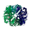
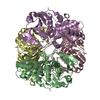
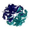
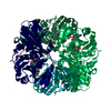
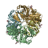
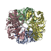
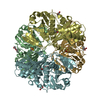
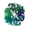
| Title | GAPDH purified from the supernatant of HEK293F cells: crystal form 1 of 4. | |||||||||
 Components Components | Glyceraldehyde-3-phosphate dehydrogenase | |||||||||
 Keywords Keywords | BIOSYNTHETIC PROTEIN / HEK293F / kifunensine / Cysteine-S-Sulfonic acid | |||||||||
| Function / homology |  Function and homology information Function and homology informationpeptidyl-cysteine S-trans-nitrosylation / Transferases; Transferring nitrogenous groups; Transferring other nitrogenous groups / negative regulation of endopeptidase activity / glyceraldehyde-3-phosphate dehydrogenase (phosphorylating) / : / aspartic-type endopeptidase inhibitor activity / glyceraldehyde-3-phosphate dehydrogenase (NAD+) (phosphorylating) activity / Gluconeogenesis / Glycolysis / canonical glycolysis ...peptidyl-cysteine S-trans-nitrosylation / Transferases; Transferring nitrogenous groups; Transferring other nitrogenous groups / negative regulation of endopeptidase activity / glyceraldehyde-3-phosphate dehydrogenase (phosphorylating) / : / aspartic-type endopeptidase inhibitor activity / glyceraldehyde-3-phosphate dehydrogenase (NAD+) (phosphorylating) activity / Gluconeogenesis / Glycolysis / canonical glycolysis / GAIT complex / peptidyl-cysteine S-nitrosylase activity / regulation of macroautophagy / defense response to fungus / positive regulation of type I interferon production / lipid droplet / positive regulation of cytokine production / glycolytic process / cellular response to type II interferon / microtubule cytoskeleton organization / NAD binding / disordered domain specific binding / NADP binding / antimicrobial humoral immune response mediated by antimicrobial peptide / neuron apoptotic process / microtubule cytoskeleton / nuclear membrane / microtubule binding / killing of cells of another organism / vesicle / positive regulation of canonical NF-kappaB signal transduction / negative regulation of translation / protein stabilization / ribonucleoprotein complex / perinuclear region of cytoplasm / extracellular exosome / membrane / identical protein binding / nucleus / plasma membrane / cytosol / cytoplasm Similarity search - Function | |||||||||
| Biological species |  Homo sapiens (human) Homo sapiens (human) | |||||||||
| Method |  X-RAY DIFFRACTION / SYNCHROTRON / MOLECULAR REPLACEMENT / Resolution: 1.525 Å X-RAY DIFFRACTION / SYNCHROTRON / MOLECULAR REPLACEMENT / Resolution: 1.525 Å | |||||||||
 Authors Authors | Roversi, P. / Lia, A. | |||||||||
| Funding support |  United Kingdom, 2items United Kingdom, 2items
| |||||||||
 Citation Citation | Journal: Wellcome Open Res / Year: 2020 Title: Partial catalytic Cys oxidation of human GAPDH to Cys-sulfonic acid. Authors: Lia, A. / Dowle, A. / Taylor, C. / Santino, A. / Roversi, P. | |||||||||
| History |
|
- Structure visualization
| Structure viewer | Molecule: MolmilJmol/JSmol |
|---|
- Downloads & links
-Download
| PDBx/mmCIF format | 6ynd.cif.gz | 989.9 KB | Display | PDBx/mmCIF format |
|---|---|---|---|---|
| PDB format | pdb6ynd.ent.gz | 835.5 KB | Display | PDB format |
| PDBx/mmJSON format | 6ynd.json.gz | Tree view | PDBx/mmJSON format | |
| Others |  Other downloads Other downloads |
-Validation report
| Arichive directory | https://data.pdbj.org/pub/pdb/validation_reports/yn/6yndftp://data.pdbj.org/pub/pdb/validation_reports/yn/6ynd | HTTPS FTP |
|---|
-Related structure data
| Related structure data | 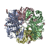 6yneC  6ynfC  6ynhC 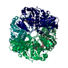 1u8fS C: citing same article ( S: Starting model for refinement |
|---|---|
| Similar structure data |


-Links
 PDBj
PDBj

- Assembly
| Deposited unit | 
| ||||||||
|---|---|---|---|---|---|---|---|---|---|
| 1 | 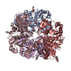
| ||||||||
| 2 | 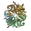
| ||||||||
| Unit cell |
|
-Components
| #1: Protein | Mass: 36179.230 Da / Num. of mol.: 8 / Mutation: C152X / Source method: isolated from a natural source Details: GAPDH with catalytic Cys partially oxidised to Cys-S-Sulfonic Acid Source: (natural) Homo sapiens (human) / Cell line: HEK293F / Organ: Kidney / Tissue: EpitheliumReferences: UniProt: P04406, glyceraldehyde-3-phosphate dehydrogenase (phosphorylating), Transferases; Transferring nitrogenous groups; Transferring other nitrogenous groups #2: Chemical | ChemComp-XPE /   Mass: 458.541 Da / Num. of mol.: 13 / Source method: obtained synthetically / Formula: C20H42O11 / Comment: precipitant*YM Mass: 458.541 Da / Num. of mol.: 13 / Source method: obtained synthetically / Formula: C20H42O11 / Comment: precipitant*YM#3: Water | ChemComp-HOH / |  Mass: 18.015 Da / Num. of mol.: 1639 / Source method: isolated from a natural source / Formula: H2O Mass: 18.015 Da / Num. of mol.: 1639 / Source method: isolated from a natural source / Formula: H2OHas ligand of interest | Y | |
|---|
-Experimental details
-Experiment
| Experiment | Method: X-RAY DIFFRACTION / Number of used crystals: 1 |
|---|
- Sample preparation
| Crystal | Density Matthews: 2.48 Å3/Da / Density % sol: 50.34 % / Description: Prism |
|---|---|
| Crystal grow | Temperature: 291 K / Method: vapor diffusion, sitting drop / pH: 8.5 Details: 0.1M MORPHEUS Amino acids solution (DL-Glutamic acid; DL-Alanine; Glycine; DL-Lysine; DL-Serine), 0.1M MORPHEUS Buffer System 3 (Tris (base); BICINE), 30% v/v MORPHEUS Precipitant Mix 1 (40% ...Details: 0.1M MORPHEUS Amino acids solution (DL-Glutamic acid; DL-Alanine; Glycine; DL-Lysine; DL-Serine), 0.1M MORPHEUS Buffer System 3 (Tris (base); BICINE), 30% v/v MORPHEUS Precipitant Mix 1 (40% v/v PEG 500* MME; 20 % w/v PEG 20000) |
-Data collection
| Diffraction | Mean temperature: 100 K / Serial crystal experiment: N |
|---|---|
| Diffraction source | Source: SYNCHROTRON / Site: Diamond / Beamline: I03 / Wavelength: 0.97622 Å |
| Detector | Type: DECTRIS EIGER2 XE 16M / Detector: PIXEL / Date: Jul 22, 2019 |
| Radiation | Monochromator: Double crystal monochromator / Protocol: SINGLE WAVELENGTH / Monochromatic (M) / Laue (L): M / Scattering type: x-ray |
| Radiation wavelength | Wavelength: 0.97622 Å / Relative weight: 1 |
| Reflection | Resolution: 1.52→140.09 Å / Num. obs: 295271 / % possible obs: 69.4 % / Redundancy: 6.9 % / Biso Wilson estimate: 23.93 Å2 / CC1/2: 0.997 / Rmerge(I) obs: 0.08 / Rpim(I) all: 0.03 / Rrim(I) all: 0.09 / Net I/σ(I): 11.7 |
| Reflection shell | Resolution: 1.52→1.71 Å / Redundancy: 6.8 % / Rmerge(I) obs: 1.071 / Mean I/σ(I) obs: 1.7 / Num. unique obs: 14763 / CC1/2: 0.608 / Rpim(I) all: 0.444 / Rrim(I) all: 1.161 / % possible all: 12.2 |
- Processing
| Software |
| ||||||||||||||||||||||||||||||||||||||||||||||||||||||||||||||||||||||||||||||||||||||||||||||||||||||||||||||||||
|---|---|---|---|---|---|---|---|---|---|---|---|---|---|---|---|---|---|---|---|---|---|---|---|---|---|---|---|---|---|---|---|---|---|---|---|---|---|---|---|---|---|---|---|---|---|---|---|---|---|---|---|---|---|---|---|---|---|---|---|---|---|---|---|---|---|---|---|---|---|---|---|---|---|---|---|---|---|---|---|---|---|---|---|---|---|---|---|---|---|---|---|---|---|---|---|---|---|---|---|---|---|---|---|---|---|---|---|---|---|---|---|---|---|---|---|
| Refinement | Method to determine structure: MOLECULAR REPLACEMENT Starting model: 1U8F Resolution: 1.525→140.09 Å / Cor.coef. Fo:Fc: 0.958 / Cor.coef. Fo:Fc free: 0.948 / SU R Cruickshank DPI: 0.135 / Cross valid method: THROUGHOUT / σ(F): 0 / SU R Blow DPI: 0.104 / SU Rfree Blow DPI: 0.095 / SU Rfree Cruickshank DPI: 0.095 Details: Initial automated water addition and positional and individual B-factor refinement were carried out in autoBUSTER. Automated non-crystallographic restraints were used throughtout, including ...Details: Initial automated water addition and positional and individual B-factor refinement were carried out in autoBUSTER. Automated non-crystallographic restraints were used throughtout, including water molecules (assigned to each chain using CCP4-Sortwater). At each catalytic Cys152 site, a 0.5:0.5 occupancy ratio mixture of Cys and Cys S-Sulfonic acid was initially modelled in Fo-Fc residual density. At each Cys152 site, occupancies for Cys and Cys S-Sulfonic acid were refined and constrained so that they sum up to 1.000 plus or minus 0.005.
| ||||||||||||||||||||||||||||||||||||||||||||||||||||||||||||||||||||||||||||||||||||||||||||||||||||||||||||||||||
| Displacement parameters | Biso mean: 27.48 Å2
| ||||||||||||||||||||||||||||||||||||||||||||||||||||||||||||||||||||||||||||||||||||||||||||||||||||||||||||||||||
| Refine analyze | Luzzati coordinate error obs: 0.22 Å | ||||||||||||||||||||||||||||||||||||||||||||||||||||||||||||||||||||||||||||||||||||||||||||||||||||||||||||||||||
| Refinement step | Cycle: LAST / Resolution: 1.525→140.09 Å
| ||||||||||||||||||||||||||||||||||||||||||||||||||||||||||||||||||||||||||||||||||||||||||||||||||||||||||||||||||
| Refine LS restraints |
| ||||||||||||||||||||||||||||||||||||||||||||||||||||||||||||||||||||||||||||||||||||||||||||||||||||||||||||||||||
| LS refinement shell | Resolution: 1.525→1.63 Å
|
