 Movie
Movie Controller

[English] 日本語
 Yorodumi
- PDB-7fiq: The crystal structure of mannose-bound beta-1,2-mannobiose phosph... -
+ Open data

- Basic information
| Entry | Database: PDB / ID: 7fiq | ||||||
|---|---|---|---|---|---|---|---|
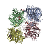
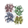
| Title | The crystal structure of mannose-bound beta-1,2-mannobiose phosphorylase from Thermoanaerobacter sp. | ||||||
 Components Components | Beta-1,2-mannobiose phosphorylase | ||||||
 Keywords Keywords | TRANSFERASE / Mannobiose / phosphorylase / thermoanaerobacter / crystallization / complex structure | ||||||
| Function / homology |  Function and homology information Function and homology informationbeta-1,2-mannobiose phosphorylase / GDP-mannose biosynthetic process / glycosyltransferase activity Similarity search - Function | ||||||
| Biological species | Thermoanaerobacter sp. | ||||||
| Method |  X-RAY DIFFRACTION / MOLECULAR REPLACEMENT / Resolution: 2.22 Å X-RAY DIFFRACTION / MOLECULAR REPLACEMENT / Resolution: 2.22 Å | ||||||
 Authors Authors | Dai, L. / Chang, Z. / Yang, J. / Liu, W. / Yang, Y. / Chen, C.-C. / Zhang, L. / Huang, J. / Sun, Y. / Guo, R.-T. | ||||||
| Funding support |  China, 1items China, 1items
| ||||||
 Citation Citation | Journal: Biochem.Biophys.Res.Commun. / Year: 2021 Title: Structural investigation of a thermostable 1,2-beta-mannobiose phosphorylase from Thermoanaerobacter sp. X-514. Authors: Dai, L. / Chang, Z. / Yang, J. / Liu, W. / Yang, Y. / Chen, C.C. / Zhang, L. / Huang, J.W. / Sun, Y. / Guo, R.T. | ||||||
| History |
|
- Structure visualization
| Structure viewer | Molecule: MolmilJmol/JSmol |
|---|
- Downloads & links
-Download
| PDBx/mmCIF format | 7fiq.cif.gz | 291.1 KB | Display | PDBx/mmCIF format |
|---|---|---|---|---|
| PDB format | pdb7fiq.ent.gz | 231.5 KB | Display | PDB format |
| PDBx/mmJSON format | 7fiq.json.gz | Tree view | PDBx/mmJSON format | |
| Others |  Other downloads Other downloads |
-Validation report
| Arichive directory | https://data.pdbj.org/pub/pdb/validation_reports/fi/7fiqftp://data.pdbj.org/pub/pdb/validation_reports/fi/7fiq | HTTPS FTP |
|---|
-Related structure data
| Related structure data | 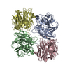 7fipC  7firC  7fisC 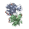 5b0rS S: Starting model for refinement C: citing same article ( |
|---|---|
| Similar structure data |


-Links
 PDBj
PDBj


- Assembly
| Deposited unit | 
| ||||||||
|---|---|---|---|---|---|---|---|---|---|
| 1 |
| ||||||||
| Unit cell |
|
-Components
-Protein / Sugars , 2 types, 8 molecules CBAD
| #1: Protein | Mass: 35856.902 Da / Num. of mol.: 4 Source method: isolated from a genetically manipulated source Source: (gene. exp.)   Thermoanaerobacter sp. (strain X514) (bacteria) Thermoanaerobacter sp. (strain X514) (bacteria)Strain: X514 / Gene: Teth514_1789 / Production host: References: UniProt: B0K2C3, beta-1,2-mannobiose phosphorylase #2: Sugar | ChemComp-MAN /  Type: D-saccharide, alpha linking / Mass: 180.156 Da / Num. of mol.: 4 / Source method: obtained synthetically / Formula: C6H12O6 / Feature type: SUBJECT OF INVESTIGATION Type: D-saccharide, alpha linking / Mass: 180.156 Da / Num. of mol.: 4 / Source method: obtained synthetically / Formula: C6H12O6 / Feature type: SUBJECT OF INVESTIGATION |
|---|
-Non-polymers , 5 types, 1358 molecules 


| #3: Chemical | ChemComp-GOL /  Mass: 92.094 Da / Num. of mol.: 1 / Source method: obtained synthetically / Formula: C3H8O3 Mass: 92.094 Da / Num. of mol.: 1 / Source method: obtained synthetically / Formula: C3H8O3 | ||||||
|---|---|---|---|---|---|---|---|
| #4: Chemical |  Mass: 150.173 Da / Num. of mol.: 2 / Source method: obtained synthetically / Formula: C6H14O4 Mass: 150.173 Da / Num. of mol.: 2 / Source method: obtained synthetically / Formula: C6H14O4#5: Chemical | ChemComp-ZN /  Mass: 65.409 Da / Num. of mol.: 17 / Source method: obtained synthetically / Formula: Zn Mass: 65.409 Da / Num. of mol.: 17 / Source method: obtained synthetically / Formula: Zn#6: Chemical | ChemComp-1PE / |  Mass: 238.278 Da / Num. of mol.: 1 / Source method: obtained synthetically / Formula: C10H22O6 / Comment: precipitant*YM Mass: 238.278 Da / Num. of mol.: 1 / Source method: obtained synthetically / Formula: C10H22O6 / Comment: precipitant*YM#7: Water | ChemComp-HOH / | Mass: 18.015 Da / Num. of mol.: 1337 / Source method: isolated from a natural source / Formula: H2O |
-Details
| Has ligand of interest | Y |
|---|
-Experimental details
-Experiment
| Experiment | Method: X-RAY DIFFRACTION / Number of used crystals: 1 |
|---|
- Sample preparation
| Crystal | Density Matthews: 2.8 Å3/Da / Density % sol: 56.12 % |
|---|---|
| Crystal grow | Temperature: 298 K / Method: evaporation / Details: 10 mM ZnCl2, 24% PEG3350, 180 mM Tris-HCl pH 7.0 |
-Data collection
| Diffraction | Mean temperature: 100 K / Serial crystal experiment: N |
|---|---|
| Diffraction source | Source: LIQUID ANODE / Type: BRUKER METALJET / Wavelength: 1.34138 Å |
| Detector | Type: BRUKER PHOTON 100 / Detector: CMOS / Date: Nov 4, 2020 |
| Radiation | Protocol: SINGLE WAVELENGTH / Monochromatic (M) / Laue (L): M / Scattering type: x-ray |
| Radiation wavelength | Wavelength: 1.34138 Å / Relative weight: 1 |
| Reflection | Resolution: 2.22→34.9 Å / Num. obs: 150222 / % possible obs: 100 % / Redundancy: 5.6 % / CC1/2: 0.992 / Rmerge(I) obs: 0.134 / Net I/σ(I): 9 |
| Reflection shell | Resolution: 2.22→2.25 Å / Rmerge(I) obs: 0.392 / Mean I/σ(I) obs: 3.1 / Num. unique obs: 3065 |
- Processing
| Software |
| ||||||||||||||||||||||||||||||||||||||||||||||||||||||||||||||||||||||||||||||||||||||||||||||||||||||||||||||||||||||||||||||||||||||||||||||||||||||||||||||||||||||||||||||||||||||||||||||||||||
|---|---|---|---|---|---|---|---|---|---|---|---|---|---|---|---|---|---|---|---|---|---|---|---|---|---|---|---|---|---|---|---|---|---|---|---|---|---|---|---|---|---|---|---|---|---|---|---|---|---|---|---|---|---|---|---|---|---|---|---|---|---|---|---|---|---|---|---|---|---|---|---|---|---|---|---|---|---|---|---|---|---|---|---|---|---|---|---|---|---|---|---|---|---|---|---|---|---|---|---|---|---|---|---|---|---|---|---|---|---|---|---|---|---|---|---|---|---|---|---|---|---|---|---|---|---|---|---|---|---|---|---|---|---|---|---|---|---|---|---|---|---|---|---|---|---|---|---|---|---|---|---|---|---|---|---|---|---|---|---|---|---|---|---|---|---|---|---|---|---|---|---|---|---|---|---|---|---|---|---|---|---|---|---|---|---|---|---|---|---|---|---|---|---|---|---|---|---|
| Refinement | Method to determine structure: MOLECULAR REPLACEMENT Starting model: 5b0r Resolution: 2.22→34.85 Å / SU ML: 0.24 / Cross valid method: THROUGHOUT / σ(F): 1.34 / Phase error: 21.27 / Stereochemistry target values: ML
| ||||||||||||||||||||||||||||||||||||||||||||||||||||||||||||||||||||||||||||||||||||||||||||||||||||||||||||||||||||||||||||||||||||||||||||||||||||||||||||||||||||||||||||||||||||||||||||||||||||
| Solvent computation | Shrinkage radii: 0.9 Å / VDW probe radii: 1.11 Å / Solvent model: FLAT BULK SOLVENT MODEL | ||||||||||||||||||||||||||||||||||||||||||||||||||||||||||||||||||||||||||||||||||||||||||||||||||||||||||||||||||||||||||||||||||||||||||||||||||||||||||||||||||||||||||||||||||||||||||||||||||||
| Displacement parameters | Biso max: 87.85 Å2 / Biso mean: 24.8869 Å2 / Biso min: 3.69 Å2 | ||||||||||||||||||||||||||||||||||||||||||||||||||||||||||||||||||||||||||||||||||||||||||||||||||||||||||||||||||||||||||||||||||||||||||||||||||||||||||||||||||||||||||||||||||||||||||||||||||||
| Refinement step | Cycle: final / Resolution: 2.22→34.85 Å
| ||||||||||||||||||||||||||||||||||||||||||||||||||||||||||||||||||||||||||||||||||||||||||||||||||||||||||||||||||||||||||||||||||||||||||||||||||||||||||||||||||||||||||||||||||||||||||||||||||||
| LS refinement shell | Refine-ID: X-RAY DIFFRACTION / Rfactor Rfree error: 0 / Total num. of bins used: 27
|
