 Movie
Movie Controller

[English] 日本語
 Yorodumi
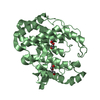
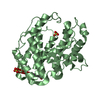
- PDB-7fi0: Crystal structure of Multi-functional Polysaccharide lyase Smlt14... -
+ Open data

- Basic information
| Entry | Database: PDB / ID: 7fi0 | |||||||||
|---|---|---|---|---|---|---|---|---|---|---|
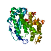
| Title | Crystal structure of Multi-functional Polysaccharide lyase Smlt1473 (WT) from Stenotrophomonas maltophilia (strain K279a) in ManA bound form at pH-5.0 | |||||||||
 Components Components | Polysaccharide lyase | |||||||||
 Keywords Keywords | LYASE / Anionic Polysaccharide lyase | |||||||||
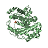
| Function / homology |  Function and homology information Function and homology informationglucuronan lyase / glucuronan lyase activity / mannuronate-specific alginate lyase / poly(beta-D-mannuronate) lyase activity / Lyases; Carbon-oxygen lyases; Acting on polysaccharides / hyaluronate lyase / hyaluronate lyase activity / polysaccharide catabolic process / cell outer membrane / periplasmic space Similarity search - Function | |||||||||
| Biological species |  Stenotrophomonas maltophilia K279a (bacteria) Stenotrophomonas maltophilia K279a (bacteria) | |||||||||
| Method |  X-RAY DIFFRACTION / MOLECULAR REPLACEMENT / Resolution: 2.31 Å X-RAY DIFFRACTION / MOLECULAR REPLACEMENT / Resolution: 2.31 Å | |||||||||
 Authors Authors | Pandey, S. / Berger, B.W. / Acharya, R. | |||||||||
| Funding support |  India, India,  United States, 2items United States, 2items
| |||||||||
 Citation Citation | Journal: J.Biol.Chem. / Year: 2021 Title: Structural insights into the mechanism of pH-selective substrate specificity of the polysaccharide lyase Smlt1473. Authors: Pandey, S. / Mahanta, P. / Berger, B.W. / Acharya, R. | |||||||||
| History |
|
- Structure visualization
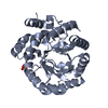
| Structure viewer | Molecule: MolmilJmol/JSmol |
|---|
- Downloads & links
-Download
| PDBx/mmCIF format | 7fi0.cif.gz | 82.3 KB | Display | PDBx/mmCIF format |
|---|---|---|---|---|
| PDB format | pdb7fi0.ent.gz | 57.9 KB | Display | PDB format |
| PDBx/mmJSON format | 7fi0.json.gz | Tree view | PDBx/mmJSON format | |
| Others |  Other downloads Other downloads |
-Validation report
| Arichive directory | https://data.pdbj.org/pub/pdb/validation_reports/fi/7fi0ftp://data.pdbj.org/pub/pdb/validation_reports/fi/7fi0 | HTTPS FTP |
|---|
-Related structure data
| Related structure data | 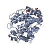 7fhuSC 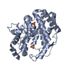 7fhvC  7fhwC 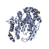 7fhxC 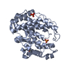 7fhyC  7fhzC  7fi1C 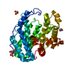 7fi2C S: Starting model for refinement C: citing same article ( |
|---|---|
| Similar structure data |


-Links
 PDBj
PDBj


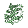
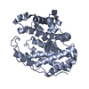
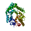
- Assembly
| Deposited unit | 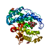
| ||||||||
|---|---|---|---|---|---|---|---|---|---|
| 1 |
| ||||||||
| Unit cell |
| ||||||||
| Components on special symmetry positions |
|
-Components
| #1: Protein | Mass: 35428.699 Da / Num. of mol.: 1 Source method: isolated from a genetically manipulated source Source: (gene. exp.) Stenotrophomonas maltophilia K279a (bacteria)Strain: K279a / Gene: Smlt1473 / Plasmid: pET28a(+) / Production host: References: UniProt: B2FHL8, Lyases; Carbon-oxygen lyases; Acting on polysaccharides |
|---|---|
| #2: Polysaccharide | beta-D-mannopyranuronic acid-(1-4)-beta-D-mannopyranuronic acid-(1-4)-beta-D-mannopyranuronic acid Source method: isolated from a genetically manipulated source |
| #3: Chemical | ChemComp-PEG /   Mass: 106.120 Da / Num. of mol.: 1 / Source method: obtained synthetically / Formula: C4H10O3 Mass: 106.120 Da / Num. of mol.: 1 / Source method: obtained synthetically / Formula: C4H10O3 |
| #4: Chemical | ChemComp-SO4 /   Mass: 96.063 Da / Num. of mol.: 1 / Source method: obtained synthetically / Formula: SO4 Mass: 96.063 Da / Num. of mol.: 1 / Source method: obtained synthetically / Formula: SO4 |
| #5: Water | ChemComp-HOH /  Mass: 18.015 Da / Num. of mol.: 173 / Source method: isolated from a natural source / Formula: H2O Mass: 18.015 Da / Num. of mol.: 173 / Source method: isolated from a natural source / Formula: H2O |
| Has ligand of interest | Y |
| Has protein modification | Y |
-Experimental details
-Experiment
| Experiment | Method: X-RAY DIFFRACTION / Number of used crystals: 1 |
|---|
- Sample preparation
| Crystal | Density Matthews: 2.89 Å3/Da / Density % sol: 57.4 % / Description: rod-shaped |
|---|---|
| Crystal grow | Temperature: 298.15 K / Method: vapor diffusion, hanging drop / pH: 5 Details: 0.2M LiSo4 Monohydrate, 0.1M Sodium acetate pH-5.0, 30% (w/v) PEG 3350 |
-Data collection
| Diffraction | Mean temperature: 100 K / Serial crystal experiment: N |
|---|---|
| Diffraction source | Source: ROTATING ANODE / Type: BRUKER AXS MICROSTAR-H / Wavelength: 1.54178 Å |
| Detector | Type: BRUKER PHOTON 100 / Detector: CMOS / Date: May 13, 2018 |
| Radiation | Protocol: SINGLE WAVELENGTH / Monochromatic (M) / Laue (L): M / Scattering type: x-ray |
| Radiation wavelength | Wavelength: 1.54178 Å / Relative weight: 1 |
| Reflection | Resolution: 2.31→29.45 Å / Num. obs: 17710 / % possible obs: 95.8 % / Redundancy: 6.39 % / CC1/2: 0.989 / Rmerge(I) obs: 0.155 / Net I/σ(I): 10.04 |
| Reflection shell | Resolution: 2.31→2.41 Å / Redundancy: 2.88 % / Rmerge(I) obs: 0.5597 / Mean I/σ(I) obs: 2.14 / Num. unique obs: 1763 / CC1/2: 0.578 / % possible all: 80.9 |
- Processing
| Software |
| |||||||||||||||||||||||||||||||||||||||||||||||||||||||||||||||||||||||||||||||||||||||||||||||||||||||||||||||||||||||||||||||||||||||||||||||||||||||||||||||||||||||||||||||||||||||||||||||||||||||||||||||||||||||||||||||||||||||
|---|---|---|---|---|---|---|---|---|---|---|---|---|---|---|---|---|---|---|---|---|---|---|---|---|---|---|---|---|---|---|---|---|---|---|---|---|---|---|---|---|---|---|---|---|---|---|---|---|---|---|---|---|---|---|---|---|---|---|---|---|---|---|---|---|---|---|---|---|---|---|---|---|---|---|---|---|---|---|---|---|---|---|---|---|---|---|---|---|---|---|---|---|---|---|---|---|---|---|---|---|---|---|---|---|---|---|---|---|---|---|---|---|---|---|---|---|---|---|---|---|---|---|---|---|---|---|---|---|---|---|---|---|---|---|---|---|---|---|---|---|---|---|---|---|---|---|---|---|---|---|---|---|---|---|---|---|---|---|---|---|---|---|---|---|---|---|---|---|---|---|---|---|---|---|---|---|---|---|---|---|---|---|---|---|---|---|---|---|---|---|---|---|---|---|---|---|---|---|---|---|---|---|---|---|---|---|---|---|---|---|---|---|---|---|---|---|---|---|---|---|---|---|---|---|---|---|---|---|---|---|---|---|
| Refinement | Method to determine structure: MOLECULAR REPLACEMENT Starting model: 7FHU Resolution: 2.31→29.402 Å / Cor.coef. Fo:Fc: 0.925 / Cor.coef. Fo:Fc free: 0.891 / Cross valid method: FREE R-VALUE / ESU R: 0.318 / ESU R Free: 0.238 Details: Hydrogens have been added in their riding positions
| |||||||||||||||||||||||||||||||||||||||||||||||||||||||||||||||||||||||||||||||||||||||||||||||||||||||||||||||||||||||||||||||||||||||||||||||||||||||||||||||||||||||||||||||||||||||||||||||||||||||||||||||||||||||||||||||||||||||
| Solvent computation | Ion probe radii: 0.8 Å / Shrinkage radii: 0.8 Å / VDW probe radii: 1.2 Å / Solvent model: MASK BULK SOLVENT | |||||||||||||||||||||||||||||||||||||||||||||||||||||||||||||||||||||||||||||||||||||||||||||||||||||||||||||||||||||||||||||||||||||||||||||||||||||||||||||||||||||||||||||||||||||||||||||||||||||||||||||||||||||||||||||||||||||||
| Displacement parameters | Biso mean: 20.624 Å2
| |||||||||||||||||||||||||||||||||||||||||||||||||||||||||||||||||||||||||||||||||||||||||||||||||||||||||||||||||||||||||||||||||||||||||||||||||||||||||||||||||||||||||||||||||||||||||||||||||||||||||||||||||||||||||||||||||||||||
| Refinement step | Cycle: LAST / Resolution: 2.31→29.402 Å
| |||||||||||||||||||||||||||||||||||||||||||||||||||||||||||||||||||||||||||||||||||||||||||||||||||||||||||||||||||||||||||||||||||||||||||||||||||||||||||||||||||||||||||||||||||||||||||||||||||||||||||||||||||||||||||||||||||||||
| Refine LS restraints |
| |||||||||||||||||||||||||||||||||||||||||||||||||||||||||||||||||||||||||||||||||||||||||||||||||||||||||||||||||||||||||||||||||||||||||||||||||||||||||||||||||||||||||||||||||||||||||||||||||||||||||||||||||||||||||||||||||||||||
| LS refinement shell | Refine-ID: X-RAY DIFFRACTION / Total num. of bins used: 20
|
