 Movie
Movie Controller


+ Open data

- Basic information
| Entry | Database: PDB / ID: 7fhl | |||||||||||||||||||||||||||
|---|---|---|---|---|---|---|---|---|---|---|---|---|---|---|---|---|---|---|---|---|---|---|---|---|---|---|---|---|
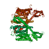
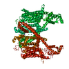
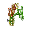
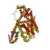
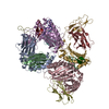
| Title | Structure of AtTPC1 with 50 mM Ca2+ | |||||||||||||||||||||||||||
 Components Components |
| |||||||||||||||||||||||||||
 Keywords Keywords | TRANSPORT PROTEIN / non-selective cation channel / dimer / vacuole | |||||||||||||||||||||||||||
| Function / homology |  Function and homology information Function and homology informationregulation of jasmonic acid biosynthetic process / seed germination / regulation of stomatal movement / plant-type vacuole / vacuole / vacuolar membrane / monoatomic ion channel complex / voltage-gated calcium channel activity / bioluminescence / calcium-mediated signaling ...regulation of jasmonic acid biosynthetic process / seed germination / regulation of stomatal movement / plant-type vacuole / vacuole / vacuolar membrane / monoatomic ion channel complex / voltage-gated calcium channel activity / bioluminescence / calcium-mediated signaling / generation of precursor metabolites and energy / calcium channel activity / calcium ion transport / calcium ion binding / Golgi apparatus / identical protein binding / plasma membrane / cytosol Similarity search - Function | |||||||||||||||||||||||||||
| Biological species |   Human respiratory syncytial virus Human respiratory syncytial virus | |||||||||||||||||||||||||||
| Method | ELECTRON MICROSCOPY / single particle reconstruction / cryo EM / Resolution: 3.1 Å | |||||||||||||||||||||||||||
 Authors Authors | Ye, F. / Xu, L. / Li, X. / Jiang, Y. / Guo, J. | |||||||||||||||||||||||||||
| Funding support |  China, 3items China, 3items
| |||||||||||||||||||||||||||
 Citation Citation | Journal: Proc Natl Acad Sci U S A / Year: 2021 Title: Voltage-gating and cytosolic Ca activation mechanisms of two-pore channel AtTPC1. Authors: Fan Ye / Lingyi Xu / Xiaoxiao Li / Weizhong Zeng / Ninghai Gan / Cheng Zhao / Wei Yang / Youxing Jiang / Jiangtao Guo /  Abstract: two-pore channel AtTPC1 is a voltage-gated, Ca-modulated, nonselective cation channel that is localized in the vacuolar membrane and responsible for generating slow vacuolar (SV) current. Under ... two-pore channel AtTPC1 is a voltage-gated, Ca-modulated, nonselective cation channel that is localized in the vacuolar membrane and responsible for generating slow vacuolar (SV) current. Under depolarizing membrane potential, cytosolic Ca activates AtTPC1 by binding at the EF-hand domain, whereas luminal Ca inhibits the channel by stabilizing the voltage-sensing domain II (VSDII) in the resting state. Here, we present 2.8 to 3.3 Å cryoelectron microscopy (cryo-EM) structures of AtTPC1 in two conformations, one in closed conformation with unbound EF-hand domain and resting VSDII and the other in a partially open conformation with Ca-bound EF-hand domain and activated VSDII. Structural comparison between the two different conformations allows us to elucidate the structural mechanisms of voltage gating, cytosolic Ca activation, and their coupling in AtTPC1. This study also provides structural insight into the general voltage-gating mechanism among voltage-gated ion channels. | |||||||||||||||||||||||||||
| History |
|
- Structure visualization
| Movie |
Movie viewer |
|---|---|
| Structure viewer | Molecule: MolmilJmol/JSmol |
- Downloads & links
-Download
| PDBx/mmCIF format | 7fhl.cif.gz | 288.5 KB | Display | PDBx/mmCIF format |
|---|---|---|---|---|
| PDB format | pdb7fhl.ent.gz | 223.6 KB | Display | PDB format |
| PDBx/mmJSON format | 7fhl.json.gz | Tree view | PDBx/mmJSON format | |
| Others |  Other downloads Other downloads |
-Validation report
| Arichive directory | https://data.pdbj.org/pub/pdb/validation_reports/fh/7fhlftp://data.pdbj.org/pub/pdb/validation_reports/fh/7fhl | HTTPS FTP |
|---|
-Related structure data
| Related structure data |  31586MC  7fhkC  7fhnC  7fhoC M: map data used to model this data C: citing same article ( |
|---|---|
| Similar structure data |


-Links
 PDBj
PDBj


- Assembly
| Deposited unit | 
|
|---|---|
| 1 |
|
-Components
| #1: Protein | Mass: 114644.297 Da / Num. of mol.: 2 Source method: isolated from a genetically manipulated source Details: The fusion protein of AtTPC1 (UNP residues 1-733), LINKER and GFP (UNP residues 2-239) Source: (gene. exp.) Human respiratory syncytial virusGene: TPC1, CCH1, FOU2, At4g03560, F9H3.19, T5L23.5 / Production host:  Homo sapiens (human) / References: UniProt: Q94KI8, UniProt: A0A5P9VSM6 Homo sapiens (human) / References: UniProt: Q94KI8, UniProt: A0A5P9VSM6#2: Protein/peptide | Mass: 1039.273 Da / Num. of mol.: 2 Source method: isolated from a genetically manipulated source Source: (gene. exp.) Homo sapiens (human)#3: Chemical | ChemComp-CA /   Mass: 40.078 Da / Num. of mol.: 4 / Source method: obtained synthetically / Formula: Ca / Feature type: SUBJECT OF INVESTIGATION Mass: 40.078 Da / Num. of mol.: 4 / Source method: obtained synthetically / Formula: Ca / Feature type: SUBJECT OF INVESTIGATIONHas ligand of interest | Y | Has protein modification | Y | Sequence details | Authors know the sequence of Chain B/D, but don't know how the coordinates align with the sequence. ...Authors know the sequence of Chain B/D, but don't know how the coordinates align with the sequence. The sequence is: CQGQDSQEKR | |
|---|
-Experimental details
-Experiment
| Experiment | Method: ELECTRON MICROSCOPY |
|---|---|
| EM experiment | Aggregation state: PARTICLE / 3D reconstruction method: single particle reconstruction |
- Sample preparation
| Component | Name: AtTPC1 homodimer / Type: COMPLEX / Entity ID: #1-#2 / Source: RECOMBINANT |
|---|---|
| Source (natural) | Organism: |
| Source (recombinant) | Organism: Homo sapiens (human) |
| Buffer solution | pH: 7.4 |
| Specimen | Embedding applied: NO / Shadowing applied: NO / Staining applied: NO / Vitrification applied: YES |
| Vitrification | Cryogen name: ETHANE |
- Electron microscopy imaging
| Experimental equipment |  Model: Titan Krios / Image courtesy: FEI Company |
|---|---|
| Microscopy | Model: FEI TITAN KRIOS |
| Electron gun | Electron source:  FIELD EMISSION GUN / Accelerating voltage: 300 kV / Illumination mode: OTHER FIELD EMISSION GUN / Accelerating voltage: 300 kV / Illumination mode: OTHER |
| Electron lens | Mode: BRIGHT FIELD |
| Image recording | Electron dose: 64 e/Å2 / Film or detector model: GATAN K2 SUMMIT (4k x 4k) |
- Processing
| Software | Name: PHENIX / Version: 1.19.2_4158: / Classification: refinement | ||||||||||||||||||||||||
|---|---|---|---|---|---|---|---|---|---|---|---|---|---|---|---|---|---|---|---|---|---|---|---|---|---|
| EM software | Name: PHENIX / Category: model refinement | ||||||||||||||||||||||||
| CTF correction | Type: PHASE FLIPPING AND AMPLITUDE CORRECTION | ||||||||||||||||||||||||
| 3D reconstruction | Resolution: 3.1 Å / Resolution method: FSC 0.143 CUT-OFF / Num. of particles: 175362 / Symmetry type: POINT | ||||||||||||||||||||||||
| Refine LS restraints |
|
