 Movie
Movie Controller


+ Open data

- Basic information
| Entry | Database: PDB / ID: 7ffe | ||||||
|---|---|---|---|---|---|---|---|
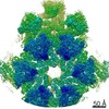
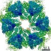
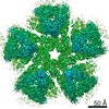
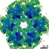
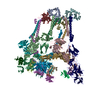
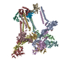
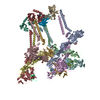
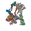
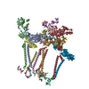
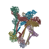
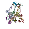
| Title | Cryo-EM structure of VEEV VLP | ||||||
 Components Components |
| ||||||
 Keywords Keywords | VIRUS LIKE PARTICLE / Virus / Receptor / Complex | ||||||
| Function / homology |  Function and homology information Function and homology informationtogavirin / T=4 icosahedral viral capsid / clathrin-dependent endocytosis of virus by host cell / symbiont-mediated suppression of host toll-like receptor signaling pathway / host cell cytoplasm / entry receptor-mediated virion attachment to host cell / symbiont-mediated suppression of host gene expression / serine-type endopeptidase activity / fusion of virus membrane with host endosome membrane / viral envelope ...togavirin / T=4 icosahedral viral capsid / clathrin-dependent endocytosis of virus by host cell / symbiont-mediated suppression of host toll-like receptor signaling pathway / host cell cytoplasm / entry receptor-mediated virion attachment to host cell / symbiont-mediated suppression of host gene expression / serine-type endopeptidase activity / fusion of virus membrane with host endosome membrane / viral envelope / host cell nucleus / host cell plasma membrane / virion membrane / structural molecule activity / proteolysis / RNA binding Similarity search - Function | ||||||
| Biological species |  Venezuelan equine encephalitis virus Venezuelan equine encephalitis virus | ||||||
| Method | ELECTRON MICROSCOPY / single particle reconstruction / cryo EM / Resolution: 3.5 Å | ||||||
 Authors Authors | Zhang, X. / Xiang, Y. / Ma, J. / Ma, B. / Huang, C. | ||||||
| Funding support | 1items
| ||||||
 Citation Citation | Journal: Nature / Year: 2021 Title: Structure of Venezuelan equine encephalitis virus with its receptor LDLRAD3. Authors: Bingting Ma / Cuiqing Huang / Jun Ma / Ye Xiang / Xinzheng Zhang /  Abstract: Venezuelan equine encephalitis virus (VEEV) is an enveloped RNA virus that causes encephalitis and potentially mortality in infected humans and equines. At present, no vaccines or drugs are available ...Venezuelan equine encephalitis virus (VEEV) is an enveloped RNA virus that causes encephalitis and potentially mortality in infected humans and equines. At present, no vaccines or drugs are available that prevent or cure diseases caused by VEEV. Low-density lipoprotein receptor class A domain-containing 3 (LDLRAD3) was recently identified as a receptor for the entry of VEEV into host cells. Here we present the cryo-electron microscopy structure of the LDLRAD3 extracellular domain 1 (LDLRAD3-D1) in complex with VEEV virus-like particles at a resolution of 3.0 Å. LDLRAD3-D1 has a cork-like structure and is inserted into clefts formed between adjacent VEEV E2-E1 heterodimers in the viral-surface trimer spikes through hydrophobic and polar contacts. Mutagenesis studies of LDLRAD3-D1 identified residues that are involved in the key interactions with VEEV. Of note, some of the LDLRAD3-D1 mutants showed a significantly increased binding affinity for VEEV, suggesting that LDLRAD3-D1 may serve as a potential scaffold for the development of inhibitors of VEEV entry. Our structures provide insights into alphavirus assembly and the binding of receptors to alphaviruses, which may guide the development of therapeutic countermeasures against alphaviruses. | ||||||
| History |
|
- Structure visualization
| Movie |
Movie viewer |
|---|---|
| Structure viewer | Molecule: MolmilJmol/JSmol |
- Downloads & links
-Download
| PDBx/mmCIF format | 7ffe.cif.gz | 727.3 KB | Display | PDBx/mmCIF format |
|---|---|---|---|---|
| PDB format | pdb7ffe.ent.gz | Display | PDB format | |
| PDBx/mmJSON format | 7ffe.json.gz | Tree view | PDBx/mmJSON format | |
| Others |  Other downloads Other downloads |
-Validation report
| Arichive directory | https://data.pdbj.org/pub/pdb/validation_reports/ff/7ffeftp://data.pdbj.org/pub/pdb/validation_reports/ff/7ffe | HTTPS FTP |
|---|
-Related structure data
| Related structure data | 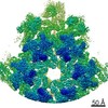 31566MC 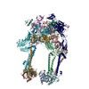 7fffC 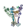 7fflC 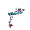 7ffnC 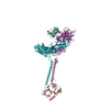 7ffoC 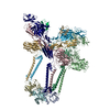 7ffqC M: map data used to model this data C: citing same article ( |
|---|---|
| Similar structure data |


-Links
 PDBj
PDBj


- Assembly
| Deposited unit | 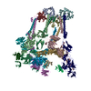
|
|---|---|
| 1 |
|
-Components
| #1: Protein | Mass: 30980.801 Da / Num. of mol.: 4 / Mutation: K64N Source method: isolated from a genetically manipulated source Source: (gene. exp.) Venezuelan equine encephalitis virus (strain TC-83)Strain: TC-83 / Production host:  Homo sapiens (human) / References: UniProt: P05674, togavirin Homo sapiens (human) / References: UniProt: P05674, togavirin#2: Protein | Mass: 47952.066 Da / Num. of mol.: 4 Source method: isolated from a genetically manipulated source Source: (gene. exp.) Venezuelan equine encephalitis virus (strain TC-83)Strain: TC-83 / Production host: Homo sapiens (human) / References: UniProt: P05674#3: Protein | Mass: 6488.601 Da / Num. of mol.: 4 Source method: isolated from a genetically manipulated source Source: (gene. exp.) Venezuelan equine encephalitis virus (strain TC-83)Strain: TC-83 / Production host: Homo sapiens (human) / References: UniProt: P05674#4: Protein | Mass: 47113.746 Da / Num. of mol.: 4 Source method: isolated from a genetically manipulated source Source: (gene. exp.) Venezuelan equine encephalitis virus (strain TC-83)Strain: TC-83 / Production host: Homo sapiens (human) / References: UniProt: P05674Has protein modification | Y | |
|---|
-Experimental details
-Experiment
| Experiment | Method: ELECTRON MICROSCOPY |
|---|---|
| EM experiment | Aggregation state: PARTICLE / 3D reconstruction method: single particle reconstruction |
- Sample preparation
| Component | Name: Venezuelan equine encephalitis virus (strain TC-83) / Type: VIRUS / Entity ID: all / Source: RECOMBINANT |
|---|---|
| Source (natural) | Organism: Venezuelan equine encephalitis virus (strain TC-83) |
| Source (recombinant) | Organism: Homo sapiens (human) |
| Details of virus | Empty: YES / Enveloped: YES / Isolate: STRAIN / Type: VIRUS-LIKE PARTICLE |
| Buffer solution | pH: 8 |
| Specimen | Embedding applied: NO / Shadowing applied: NO / Staining applied: NO / Vitrification applied: YES |
| Vitrification | Cryogen name: ETHANE |
- Electron microscopy imaging
| Experimental equipment |  Model: Talos Arctica / Image courtesy: FEI Company |
|---|---|
| Microscopy | Model: FEI TECNAI ARCTICA |
| Electron gun | Electron source:  FIELD EMISSION GUN / Accelerating voltage: 200 kV / Illumination mode: FLOOD BEAM FIELD EMISSION GUN / Accelerating voltage: 200 kV / Illumination mode: FLOOD BEAM |
| Electron lens | Mode: BRIGHT FIELD |
| Image recording | Electron dose: 40 e/Å2 / Film or detector model: GATAN K2 SUMMIT (4k x 4k) |
- Processing
| CTF correction | Type: PHASE FLIPPING AND AMPLITUDE CORRECTION |
|---|---|
| 3D reconstruction | Resolution: 3.5 Å / Resolution method: FSC 0.143 CUT-OFF / Num. of particles: 344824 / Symmetry type: POINT |
