 Movie
Movie Controller

[English] 日本語
 Yorodumi
- PDB-7fe1: Crystal structure of GH92 alpha-1,2-mannosidase from Enterococcus... -
+ Open data

- Basic information
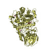
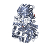
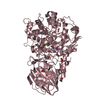
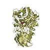
| Entry | Database: PDB / ID: 7fe1 | ||||||
|---|---|---|---|---|---|---|---|
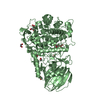
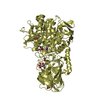
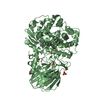
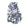
| Title | Crystal structure of GH92 alpha-1,2-mannosidase from Enterococcus faecalis ATCC 10100 in complex with methyl alpha-1,2-C-mannobioside | ||||||
 Components Components | Alpha-1,2-mannosidase | ||||||
 Keywords Keywords | HYDROLASE / Glycoside hydrolase / GH92 / Inhibitor / Carbohydrate / N-glycan | ||||||
| Function / homology | Beta-galactosidase; Chain A, domain 5 - #10 / Beta-galactosidase; Chain A, domain 5 / Distorted Sandwich / Mainly Beta / methyl 2-deoxy-2-methyl-alpha-D-mannopyranoside / ACETIC ACID / alpha-D-mannopyranose / :  Function and homology information Function and homology information | ||||||
| Biological species |  Enterococcus faecalis ATCC 10100 (bacteria) Enterococcus faecalis ATCC 10100 (bacteria) | ||||||
| Method |  X-RAY DIFFRACTION / SYNCHROTRON / MOLECULAR REPLACEMENT / Resolution: 1.72 Å X-RAY DIFFRACTION / SYNCHROTRON / MOLECULAR REPLACEMENT / Resolution: 1.72 Å | ||||||
 Authors Authors | Miyazaki, T. / Alonso-Gil, S. | ||||||
| Funding support |  Japan, 1items Japan, 1items
| ||||||
 Citation Citation | Journal: Chemistry / Year: 2022 Title: Unlocking the Hydrolytic Mechanism of GH92 alpha-1,2-Mannosidases: Computation Inspires the use of C-Glycosides as Michaelis Complex Mimics. Authors: Alonso-Gil, S. / Parkan, K. / Kaminsky, J. / Pohl, R. / Miyazaki, T. | ||||||
| History |
|
- Structure visualization
| Structure viewer | Molecule: MolmilJmol/JSmol |
|---|
- Downloads & links
-Download
| PDBx/mmCIF format | 7fe1.cif.gz | 659.1 KB | Display | PDBx/mmCIF format |
|---|---|---|---|---|
| PDB format | pdb7fe1.ent.gz | 515.4 KB | Display | PDB format |
| PDBx/mmJSON format | 7fe1.json.gz | Tree view | PDBx/mmJSON format | |
| Others |  Other downloads Other downloads |
-Validation report
| Arichive directory | https://data.pdbj.org/pub/pdb/validation_reports/fe/7fe1ftp://data.pdbj.org/pub/pdb/validation_reports/fe/7fe1 | HTTPS FTP |
|---|
-Related structure data
| Related structure data |  7fe2C 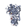 6dwoS S: Starting model for refinement C: citing same article ( |
|---|---|
| Similar structure data |

-Links
 PDBj
PDBj


- Assembly
| Deposited unit | 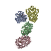
| ||||||||
|---|---|---|---|---|---|---|---|---|---|
| 1 | 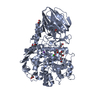
| ||||||||
| 2 | 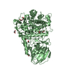
| ||||||||
| 3 | 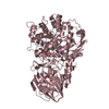
| ||||||||
| 4 | 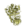
| ||||||||
| Unit cell |
|
-Components
-Protein , 1 types, 4 molecules ABCD
| #1: Protein | Mass: 82734.070 Da / Num. of mol.: 4 Source method: isolated from a genetically manipulated source Source: (gene. exp.) Enterococcus faecalis ATCC 10100 (bacteria)Gene: WOW_02008 / Plasmid: pET21a / Production host: |
|---|
-Sugars , 2 types, 8 molecules 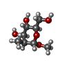

| #2: Sugar | ChemComp-5II /  Type: D-saccharide, alpha linking / Mass: 192.210 Da / Num. of mol.: 4 Type: D-saccharide, alpha linking / Mass: 192.210 Da / Num. of mol.: 4Source method: isolated from a genetically manipulated source Formula: C8H16O5 / Feature type: SUBJECT OF INVESTIGATION #3: Sugar | ChemComp-MAN /  Type: D-saccharide, alpha linking / Mass: 180.156 Da / Num. of mol.: 4 Type: D-saccharide, alpha linking / Mass: 180.156 Da / Num. of mol.: 4Source method: isolated from a genetically manipulated source Formula: C6H12O6 / Feature type: SUBJECT OF INVESTIGATION |
|---|
-Non-polymers , 5 types, 2788 molecules 


| #4: Chemical | ChemComp-CA /  Mass: 40.078 Da / Num. of mol.: 4 / Source method: obtained synthetically / Formula: Ca Mass: 40.078 Da / Num. of mol.: 4 / Source method: obtained synthetically / Formula: Ca#5: Chemical | ChemComp-NA /  Mass: 22.990 Da / Num. of mol.: 4 / Source method: obtained synthetically / Formula: Na Mass: 22.990 Da / Num. of mol.: 4 / Source method: obtained synthetically / Formula: Na#6: Chemical |  Mass: 60.052 Da / Num. of mol.: 2 / Source method: obtained synthetically / Formula: C2H4O2 Mass: 60.052 Da / Num. of mol.: 2 / Source method: obtained synthetically / Formula: C2H4O2#7: Chemical | ChemComp-EDO /  Mass: 62.068 Da / Num. of mol.: 18 / Source method: obtained synthetically / Formula: C2H6O2 Mass: 62.068 Da / Num. of mol.: 18 / Source method: obtained synthetically / Formula: C2H6O2#8: Water | ChemComp-HOH / | Mass: 18.015 Da / Num. of mol.: 2760 / Source method: isolated from a natural source / Formula: H2O |
|---|
-Details
| Has ligand of interest | Y |
|---|---|
| Nonpolymer details | methyl alpha-1,2-C-mannobioside is compounded from MAN and 5II. |
-Experimental details
-Experiment
| Experiment | Method: X-RAY DIFFRACTION / Number of used crystals: 1 |
|---|
- Sample preparation
| Crystal | Density Matthews: 2.7 Å3/Da / Density % sol: 54.5 % |
|---|---|
| Crystal grow | Temperature: 293 K / Method: vapor diffusion, hanging drop Details: 100 mM sodium citrate, pH 5.0-5.6, 100 mM magnesium acetate, 200 mM ammonium sulfate, 5% PEG 20000 PH range: 5.0-5.6 |
-Data collection
| Diffraction | Mean temperature: 100 K / Serial crystal experiment: N |
|---|---|
| Diffraction source | Source: SYNCHROTRON / Site: Photon Factory / Beamline: AR-NW12A / Wavelength: 1 Å |
| Detector | Type: DECTRIS PILATUS3 S 2M / Detector: PIXEL / Date: Mar 7, 2021 |
| Radiation | Protocol: SINGLE WAVELENGTH / Monochromatic (M) / Laue (L): M / Scattering type: x-ray |
| Radiation wavelength | Wavelength: 1 Å / Relative weight: 1 |
| Reflection | Resolution: 1.72→50 Å / Num. obs: 375878 / % possible obs: 100 % / Redundancy: 13.6 % / CC1/2: 0.999 / Rmerge(I) obs: 0.121 / Rpim(I) all: 0.034 / Rrim(I) all: 0.125 / Net I/σ(I): 17.3 |
| Reflection shell | Resolution: 1.72→1.81 Å / Redundancy: 13.5 % / Rmerge(I) obs: 1.168 / Mean I/σ(I) obs: 2.5 / Num. unique obs: 54650 / CC1/2: 0.759 / Rpim(I) all: 0.327 / Rrim(I) all: 1.214 / % possible all: 100 |
- Processing
| Software |
| ||||||||||||||||||||||||||||||||||||||||||||||||||||||||||||||||||||||||||||||||||||||||||||||||||||||||||||||||||||||||||||||||||||||||||||||||||||||||||||||||
|---|---|---|---|---|---|---|---|---|---|---|---|---|---|---|---|---|---|---|---|---|---|---|---|---|---|---|---|---|---|---|---|---|---|---|---|---|---|---|---|---|---|---|---|---|---|---|---|---|---|---|---|---|---|---|---|---|---|---|---|---|---|---|---|---|---|---|---|---|---|---|---|---|---|---|---|---|---|---|---|---|---|---|---|---|---|---|---|---|---|---|---|---|---|---|---|---|---|---|---|---|---|---|---|---|---|---|---|---|---|---|---|---|---|---|---|---|---|---|---|---|---|---|---|---|---|---|---|---|---|---|---|---|---|---|---|---|---|---|---|---|---|---|---|---|---|---|---|---|---|---|---|---|---|---|---|---|---|---|---|---|---|
| Refinement | Method to determine structure: MOLECULAR REPLACEMENT Starting model: 6DWO Resolution: 1.72→48.621 Å / Cor.coef. Fo:Fc: 0.973 / Cor.coef. Fo:Fc free: 0.965 / SU B: 1.872 / SU ML: 0.058 / Cross valid method: FREE R-VALUE / ESU R: 0.084 / ESU R Free: 0.081 Details: Hydrogens have been added in their riding positions
| ||||||||||||||||||||||||||||||||||||||||||||||||||||||||||||||||||||||||||||||||||||||||||||||||||||||||||||||||||||||||||||||||||||||||||||||||||||||||||||||||
| Solvent computation | Ion probe radii: 0.8 Å / Shrinkage radii: 0.8 Å / VDW probe radii: 1.2 Å / Solvent model: BABINET MODEL PLUS MASK | ||||||||||||||||||||||||||||||||||||||||||||||||||||||||||||||||||||||||||||||||||||||||||||||||||||||||||||||||||||||||||||||||||||||||||||||||||||||||||||||||
| Displacement parameters | Biso mean: 23.049 Å2
| ||||||||||||||||||||||||||||||||||||||||||||||||||||||||||||||||||||||||||||||||||||||||||||||||||||||||||||||||||||||||||||||||||||||||||||||||||||||||||||||||
| Refinement step | Cycle: LAST / Resolution: 1.72→48.621 Å
| ||||||||||||||||||||||||||||||||||||||||||||||||||||||||||||||||||||||||||||||||||||||||||||||||||||||||||||||||||||||||||||||||||||||||||||||||||||||||||||||||
| Refine LS restraints |
| ||||||||||||||||||||||||||||||||||||||||||||||||||||||||||||||||||||||||||||||||||||||||||||||||||||||||||||||||||||||||||||||||||||||||||||||||||||||||||||||||
| LS refinement shell |
|
