 Movie
Movie Controller

[English] 日本語
 Yorodumi
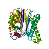
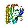
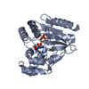
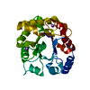
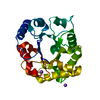
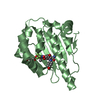
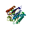
- PDB-7fds: High resolution crystal structure of LpqH from Mycobacterium tube... -
+ Open data

- Basic information
| Entry | Database: PDB / ID: 7fds | ||||||
|---|---|---|---|---|---|---|---|
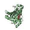
| Title | High resolution crystal structure of LpqH from Mycobacterium tuberculosis | ||||||
 Components Components | Lipoprotein LpqH | ||||||
 Keywords Keywords | LIPID BINDING PROTEIN | ||||||
| Function / homology |  Function and homology information Function and homology informationsymbiont-mediated perturbation of host immune response / bacterial extracellular vesicle / biological process involved in interaction with host / peptidoglycan-based cell wall / host cell cytoplasm / defense response to bacterium / host cell surface receptor binding / cell surface / plasma membrane Similarity search - Function | ||||||
| Biological species |  Mycobacterium tuberculosis H37Rv (bacteria) Mycobacterium tuberculosis H37Rv (bacteria) | ||||||
| Method |  X-RAY DIFFRACTION / SYNCHROTRON / MOLECULAR REPLACEMENT / Resolution: 1.258 Å X-RAY DIFFRACTION / SYNCHROTRON / MOLECULAR REPLACEMENT / Resolution: 1.258 Å | ||||||
 Authors Authors | Kundapura, S.V. / Chatterjee, S. / Samanta, D. / Ramagopal, U.A. | ||||||
 Citation Citation | Journal: Int.J.Biol.Macromol. / Year: 2022 Title: High-resolution crystal structure of LpqH, an immunomodulatory surface lipoprotein of Mycobacterium tuberculosis reveals a distinct fold and a conserved cleft on its surface. Authors: Chatterjee, S. / Kundapura, S.V. / Basak, A.J. / Mukherjee, D. / Dash, S. / Ganguli, N. / Das, A.K. / Mukherjee, G. / Samanta, D. / Ramagopal, U.A. | ||||||
| History |
|
- Structure visualization
| Structure viewer | Molecule: MolmilJmol/JSmol |
|---|
- Downloads & links
-Download
| PDBx/mmCIF format | 7fds.cif.gz | 95.5 KB | Display | PDBx/mmCIF format |
|---|---|---|---|---|
| PDB format | pdb7fds.ent.gz | 69.5 KB | Display | PDB format |
| PDBx/mmJSON format | 7fds.json.gz | Tree view | PDBx/mmJSON format | |
| Others |  Other downloads Other downloads |
-Validation report
| Arichive directory | https://data.pdbj.org/pub/pdb/validation_reports/fd/7fdsftp://data.pdbj.org/pub/pdb/validation_reports/fd/7fds | HTTPS FTP |
|---|
-Related structure data
| Related structure data | 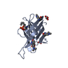 4zjmS S: Starting model for refinement |
|---|---|
| Similar structure data |


-Links
 PDBj
PDBj


- Assembly
| Deposited unit | 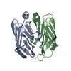
| ||||||||
|---|---|---|---|---|---|---|---|---|---|
| 1 |
| ||||||||
| Unit cell |
|
-Components
| #1: Protein | Mass: 12792.177 Da / Num. of mol.: 2 Source method: isolated from a genetically manipulated source Source: (gene. exp.) Mycobacterium tuberculosis H37Rv (bacteria)Strain: H37Rv / Gene: lpqH / Production host: #2: Water | ChemComp-HOH / |  Mass: 18.015 Da / Num. of mol.: 116 / Source method: isolated from a natural source / Formula: H2O Mass: 18.015 Da / Num. of mol.: 116 / Source method: isolated from a natural source / Formula: H2OHas protein modification | Y | |
|---|
-Experimental details
-Experiment
| Experiment | Method: X-RAY DIFFRACTION / Number of used crystals: 1 |
|---|
- Sample preparation
| Crystal | Density Matthews: 1.85 Å3/Da / Density % sol: 33.41 % |
|---|---|
| Crystal grow | Temperature: 298 K / Method: vapor diffusion, sitting drop / pH: 6.3 / Details: 0.2M Ammonium sulphate, 20% PEG 3350 |
-Data collection
| Diffraction | Mean temperature: 100 K / Serial crystal experiment: N |
|---|---|
| Diffraction source | Source: SYNCHROTRON / Site: ELETTRA  / Beamline: 11.2C / Wavelength: 1 Å / Beamline: 11.2C / Wavelength: 1 Å |
| Detector | Type: DECTRIS PILATUS 6M / Detector: PIXEL / Date: Feb 15, 2021 |
| Radiation | Protocol: SINGLE WAVELENGTH / Monochromatic (M) / Laue (L): M / Scattering type: x-ray |
| Radiation wavelength | Wavelength: 1 Å / Relative weight: 1 |
| Reflection | Resolution: 1.258→28.557 Å / Num. obs: 49917 / % possible obs: 99.1 % / Redundancy: 4.5 % / CC1/2: 0.997 / Rmerge(I) obs: 0.061 / Rpim(I) all: 0.047 / Rrim(I) all: 0.078 / Rsym value: 0.067 / Net I/σ(I): 10.1 |
| Reflection shell | Resolution: 1.258→1.28 Å / Rmerge(I) obs: 0.502 / Num. unique obs: 2627 / CC1/2: 0.811 / Rpim(I) all: 0.407 / Rrim(I) all: 0.649 / Rsym value: 0.58 / % possible all: 98 |
- Processing
| Software |
| ||||||||||||||||||||||||||||||||||||||||||||||||||||||||||||||||||||||||||||||||||||||||||||||||||||||||||||||||||||||||||||||||||||||||||||||||||||||
|---|---|---|---|---|---|---|---|---|---|---|---|---|---|---|---|---|---|---|---|---|---|---|---|---|---|---|---|---|---|---|---|---|---|---|---|---|---|---|---|---|---|---|---|---|---|---|---|---|---|---|---|---|---|---|---|---|---|---|---|---|---|---|---|---|---|---|---|---|---|---|---|---|---|---|---|---|---|---|---|---|---|---|---|---|---|---|---|---|---|---|---|---|---|---|---|---|---|---|---|---|---|---|---|---|---|---|---|---|---|---|---|---|---|---|---|---|---|---|---|---|---|---|---|---|---|---|---|---|---|---|---|---|---|---|---|---|---|---|---|---|---|---|---|---|---|---|---|---|---|---|---|
| Refinement | Method to determine structure: MOLECULAR REPLACEMENT Starting model: 4ZJM Resolution: 1.258→28.557 Å / Cor.coef. Fo:Fc: 0.977 / Cor.coef. Fo:Fc free: 0.969 / SU B: 2.031 / SU ML: 0.038 / Cross valid method: FREE R-VALUE / ESU R: 0.043 / ESU R Free: 0.044 Details: Hydrogens have been added in their riding positions
| ||||||||||||||||||||||||||||||||||||||||||||||||||||||||||||||||||||||||||||||||||||||||||||||||||||||||||||||||||||||||||||||||||||||||||||||||||||||
| Solvent computation | Ion probe radii: 0.8 Å / Shrinkage radii: 0.8 Å / VDW probe radii: 1.2 Å / Solvent model: MASK BULK SOLVENT | ||||||||||||||||||||||||||||||||||||||||||||||||||||||||||||||||||||||||||||||||||||||||||||||||||||||||||||||||||||||||||||||||||||||||||||||||||||||
| Displacement parameters | Biso mean: 19.54 Å2
| ||||||||||||||||||||||||||||||||||||||||||||||||||||||||||||||||||||||||||||||||||||||||||||||||||||||||||||||||||||||||||||||||||||||||||||||||||||||
| Refinement step | Cycle: LAST / Resolution: 1.258→28.557 Å
| ||||||||||||||||||||||||||||||||||||||||||||||||||||||||||||||||||||||||||||||||||||||||||||||||||||||||||||||||||||||||||||||||||||||||||||||||||||||
| Refine LS restraints |
| ||||||||||||||||||||||||||||||||||||||||||||||||||||||||||||||||||||||||||||||||||||||||||||||||||||||||||||||||||||||||||||||||||||||||||||||||||||||
| LS refinement shell |
|
