 Movie
Movie Controller

[English] 日本語
 Yorodumi
- PDB-7f8c: Crystal structure of rRNA methyltransferase Erm38 in complex with... -
+ Open data

- Basic information
| Entry | Database: PDB / ID: 7f8c | ||||||
|---|---|---|---|---|---|---|---|
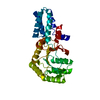
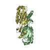
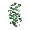
| Title | Crystal structure of rRNA methyltransferase Erm38 in complex with sinefungin | ||||||
 Components Components | Erm(38) | ||||||
 Keywords Keywords | TRANSFERASE / erythromycin resistance methyltransferase / methyltransferase / sinefungin | ||||||
| Function / homology |  Function and homology information Function and homology informationrRNA (adenine-N6,N6-)-dimethyltransferase activity / RNA binding / cytosol Similarity search - Function | ||||||
| Biological species |  Mycolicibacterium smegmatis (bacteria) Mycolicibacterium smegmatis (bacteria) | ||||||
| Method |  X-RAY DIFFRACTION / SYNCHROTRON / MOLECULAR REPLACEMENT / Resolution: 2.248 Å X-RAY DIFFRACTION / SYNCHROTRON / MOLECULAR REPLACEMENT / Resolution: 2.248 Å | ||||||
 Authors Authors | Goh, B.C. / Lescar, J. | ||||||
| Funding support |  Singapore, 1items Singapore, 1items
| ||||||
 Citation Citation | Journal: J.Biol.Chem. / Year: 2022 Title: Crystal structure and functional analysis of mycobacterial erythromycin resistance methyltransferase Erm38 reveals its RNA-binding site. Authors: Goh, B.C. / Xiang, X. / Lescar, J. / Dedon, P.C. | ||||||
| History |
|
- Structure visualization
| Structure viewer | Molecule: MolmilJmol/JSmol |
|---|
- Downloads & links
-Download
| PDBx/mmCIF format | 7f8c.cif.gz | 65.9 KB | Display | PDBx/mmCIF format |
|---|---|---|---|---|
| PDB format | pdb7f8c.ent.gz | 45.8 KB | Display | PDB format |
| PDBx/mmJSON format | 7f8c.json.gz | Tree view | PDBx/mmJSON format | |
| Others |  Other downloads Other downloads |
-Validation report
| Summary document | 7f8c_validation.pdf.gz | 763.8 KB | Display | wwPDB validaton report |
|---|---|---|---|---|
| Full document | 7f8c_full_validation.pdf.gz | 763.9 KB | Display | |
| Data in XML | 7f8c_validation.xml.gz | 12.6 KB | Display | |
| Data in CIF | 7f8c_validation.cif.gz | 17.5 KB | Display | |
| Arichive directory | https://data.pdbj.org/pub/pdb/validation_reports/f8/7f8cftp://data.pdbj.org/pub/pdb/validation_reports/f8/7f8c | HTTPS FTP |
-Related structure data
| Related structure data | 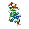 7f8aSC 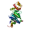 7f8bC S: Starting model for refinement C: citing same article ( |
|---|---|
| Similar structure data |


-Links
 PDBj
PDBj
- Assembly
| Deposited unit | 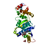
| ||||||||
|---|---|---|---|---|---|---|---|---|---|
| 1 |
| ||||||||
| Unit cell |
|
-Components
| #1: Protein | Mass: 28923.498 Da / Num. of mol.: 1 Source method: isolated from a genetically manipulated source Source: (gene. exp.) Mycolicibacterium smegmatis (bacteria) / Gene: erm(38) / Production host: |
|---|---|
| #2: Chemical | ChemComp-SFG /   Mass: 381.387 Da / Num. of mol.: 1 / Source method: obtained synthetically / Formula: C15H23N7O5 / Feature type: SUBJECT OF INVESTIGATION Mass: 381.387 Da / Num. of mol.: 1 / Source method: obtained synthetically / Formula: C15H23N7O5 / Feature type: SUBJECT OF INVESTIGATION |
| #3: Chemical | ChemComp-SIN /   Mass: 118.088 Da / Num. of mol.: 1 / Source method: obtained synthetically / Formula: C4H6O4 Mass: 118.088 Da / Num. of mol.: 1 / Source method: obtained synthetically / Formula: C4H6O4 |
| #4: Water | ChemComp-HOH /  Mass: 18.015 Da / Num. of mol.: 136 / Source method: isolated from a natural source / Formula: H2O Mass: 18.015 Da / Num. of mol.: 136 / Source method: isolated from a natural source / Formula: H2O |
| Has ligand of interest | Y |
-Experimental details
-Experiment
| Experiment | Method: X-RAY DIFFRACTION / Number of used crystals: 1 |
|---|
- Sample preparation
| Crystal | Density Matthews: 2.67 Å3/Da / Density % sol: 53.95 % |
|---|---|
| Crystal grow | Temperature: 293 K / Method: vapor diffusion, hanging drop / pH: 7 / Details: 1.0 M succinic acid pH 7, 5% glycerol |
-Data collection
| Diffraction | Mean temperature: 100 K / Serial crystal experiment: N | ||||||||||||||||||||||||||||||
|---|---|---|---|---|---|---|---|---|---|---|---|---|---|---|---|---|---|---|---|---|---|---|---|---|---|---|---|---|---|---|---|
| Diffraction source | Source: SYNCHROTRON / Site: SOLEIL  / Beamline: PROXIMA 2 / Wavelength: 0.98 Å / Beamline: PROXIMA 2 / Wavelength: 0.98 Å | ||||||||||||||||||||||||||||||
| Detector | Type: DECTRIS EIGER X 9M / Detector: PIXEL / Date: Feb 12, 2018 | ||||||||||||||||||||||||||||||
| Radiation | Protocol: SINGLE WAVELENGTH / Monochromatic (M) / Laue (L): M / Scattering type: x-ray | ||||||||||||||||||||||||||||||
| Radiation wavelength | Wavelength: 0.98 Å / Relative weight: 1 | ||||||||||||||||||||||||||||||
| Reflection | Resolution: 2.248→48.47 Å / Num. obs: 15503 / % possible obs: 99.8 % / Redundancy: 25.9 % / Biso Wilson estimate: 48.43 Å2 / CC1/2: 1 / Rmerge(I) obs: 0.079 / Rpim(I) all: 0.016 / Rrim(I) all: 0.08 / Net I/σ(I): 31.3 | ||||||||||||||||||||||||||||||
| Reflection shell | Diffraction-ID: 1
|
- Processing
| Software |
| ||||||||||||||||||||||||||||||||||||||||||||||||||||||||||||||||||||||||||||||||||||||||||||||||||||||||||||
|---|---|---|---|---|---|---|---|---|---|---|---|---|---|---|---|---|---|---|---|---|---|---|---|---|---|---|---|---|---|---|---|---|---|---|---|---|---|---|---|---|---|---|---|---|---|---|---|---|---|---|---|---|---|---|---|---|---|---|---|---|---|---|---|---|---|---|---|---|---|---|---|---|---|---|---|---|---|---|---|---|---|---|---|---|---|---|---|---|---|---|---|---|---|---|---|---|---|---|---|---|---|---|---|---|---|---|---|---|---|
| Refinement | Method to determine structure: MOLECULAR REPLACEMENT Starting model: 7F8A Resolution: 2.248→48.47 Å / Cor.coef. Fo:Fc: 0.93 / Cor.coef. Fo:Fc free: 0.922 / SU R Cruickshank DPI: 0.257 / Cross valid method: THROUGHOUT / σ(F): 0 / SU R Blow DPI: 0.279 / SU Rfree Blow DPI: 0.217 / SU Rfree Cruickshank DPI: 0.211
| ||||||||||||||||||||||||||||||||||||||||||||||||||||||||||||||||||||||||||||||||||||||||||||||||||||||||||||
| Displacement parameters | Biso max: 93.44 Å2 / Biso mean: 43.25 Å2 / Biso min: 26.27 Å2
| ||||||||||||||||||||||||||||||||||||||||||||||||||||||||||||||||||||||||||||||||||||||||||||||||||||||||||||
| Refine analyze | Luzzati coordinate error obs: 0.29 Å | ||||||||||||||||||||||||||||||||||||||||||||||||||||||||||||||||||||||||||||||||||||||||||||||||||||||||||||
| Refinement step | Cycle: final / Resolution: 2.248→48.47 Å
| ||||||||||||||||||||||||||||||||||||||||||||||||||||||||||||||||||||||||||||||||||||||||||||||||||||||||||||
| Refine LS restraints |
| ||||||||||||||||||||||||||||||||||||||||||||||||||||||||||||||||||||||||||||||||||||||||||||||||||||||||||||
| LS refinement shell | Resolution: 2.25→2.27 Å / Rfactor Rfree error: 0 / Total num. of bins used: 39
|
