 Movie
Movie Controller

[English] 日本語
 Yorodumi
- PDB-7f3i: Crystal structure of human YBX2 CSD in complex with m5C RNA in sp... -
+ Open data

- Basic information
| Entry | Database: PDB / ID: 7f3i | ||||||
|---|---|---|---|---|---|---|---|
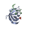
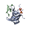
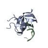
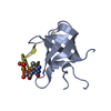
| Title | Crystal structure of human YBX2 CSD in complex with m5C RNA in space group P212121 | ||||||
 Components Components |
| ||||||
 Keywords Keywords | RNA BINDING PROTEIN/RNA / Complex / protein-RNA / YBX / Cold-shock domain / RNA BINDING PROTEIN / RNA BINDING PROTEIN-RNA complex | ||||||
| Function / homology |  Function and homology information Function and homology informationtranslational attenuation / oocyte development / positive regulation of cold-induced thermogenesis / regulation of gene expression / spermatogenesis / nucleic acid binding / transcription by RNA polymerase II / DNA binding / RNA binding / nucleus / cytoplasm Similarity search - Function | ||||||
| Biological species |  Homo sapiens (human) Homo sapiens (human) | ||||||
| Method |  X-RAY DIFFRACTION / SYNCHROTRON / MOLECULAR REPLACEMENT / Resolution: 2.25 Å X-RAY DIFFRACTION / SYNCHROTRON / MOLECULAR REPLACEMENT / Resolution: 2.25 Å | ||||||
 Authors Authors | Zhang, Y. / Huang, Y. | ||||||
| Funding support |  China, 1items China, 1items
| ||||||
 Citation Citation | Journal: To Be Published Title: Crystal structure of human YBX2 CSD in complex with m5C RNA in space group P212121 Authors: Zhang, Y. / Huang, Y. | ||||||
| History |
|
- Structure visualization
| Structure viewer | Molecule: MolmilJmol/JSmol |
|---|
- Downloads & links
-Download
| PDBx/mmCIF format | 7f3i.cif.gz | 90.9 KB | Display | PDBx/mmCIF format |
|---|---|---|---|---|
| PDB format | pdb7f3i.ent.gz | 54.8 KB | Display | PDB format |
| PDBx/mmJSON format | 7f3i.json.gz | Tree view | PDBx/mmJSON format | |
| Others |  Other downloads Other downloads |
-Validation report
| Summary document | 7f3i_validation.pdf.gz | 469.7 KB | Display | wwPDB validaton report |
|---|---|---|---|---|
| Full document | 7f3i_full_validation.pdf.gz | 474.7 KB | Display | |
| Data in XML | 7f3i_validation.xml.gz | 15.4 KB | Display | |
| Data in CIF | 7f3i_validation.cif.gz | 21 KB | Display | |
| Arichive directory | https://data.pdbj.org/pub/pdb/validation_reports/f3/7f3iftp://data.pdbj.org/pub/pdb/validation_reports/f3/7f3i | HTTPS FTP |
-Related structure data
| Related structure data | 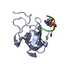 6a6jS S: Starting model for refinement |
|---|---|
| Similar structure data |

-Links
 PDBj
PDBj- Assembly
| Deposited unit | 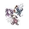
| ||||||||||||
|---|---|---|---|---|---|---|---|---|---|---|---|---|---|
| 1 |
| ||||||||||||
| Unit cell |
| ||||||||||||
| Noncrystallographic symmetry (NCS) | NCS oper:
|
-Components
| #1: Protein | Mass: 10205.441 Da / Num. of mol.: 3 / Mutation: I92T, Q93K Source method: isolated from a genetically manipulated source Source: (gene. exp.) Homo sapiens (human) / Gene: YBX2, CSDA3, MSY2 / Production host:  #2: RNA chain | Mass: 1550.996 Da / Num. of mol.: 2 / Source method: obtained synthetically / Source: (synth.) Homo sapiens (human)#3: Water | ChemComp-HOH / |  Mass: 18.015 Da / Num. of mol.: 142 / Source method: isolated from a natural source / Formula: H2O Mass: 18.015 Da / Num. of mol.: 142 / Source method: isolated from a natural source / Formula: H2OHas ligand of interest | Y | |
|---|
-Experimental details
-Experiment
| Experiment | Method: X-RAY DIFFRACTION / Number of used crystals: 1 |
|---|
- Sample preparation
| Crystal | Density Matthews: 2.3 Å3/Da / Density % sol: 46.47 % |
|---|---|
| Crystal grow | Temperature: 290 K / Method: vapor diffusion, hanging drop / Details: 0.1 M Imidazole, 20% (w/v) PEG 8000, 3% (v/v) MPD |
-Data collection
| Diffraction | Mean temperature: 100 K / Serial crystal experiment: N |
|---|---|
| Diffraction source | Source: SYNCHROTRON / Site: SSRF / Beamline: BL18U1 / Wavelength: 0.97915 Å |
| Detector | Type: DECTRIS PILATUS 6M / Detector: PIXEL / Date: Jul 17, 2020 |
| Radiation | Protocol: SINGLE WAVELENGTH / Monochromatic (M) / Laue (L): M / Scattering type: x-ray |
| Radiation wavelength | Wavelength: 0.97915 Å / Relative weight: 1 |
| Reflection | Resolution: 2.25→30 Å / Num. obs: 15180 / % possible obs: 99.9 % / Redundancy: 10.2 % / Biso Wilson estimate: 29.94 Å2 / Rmerge(I) obs: 0.124 / Net I/σ(I): 18.2 |
| Reflection shell | Resolution: 2.25→2.29 Å / Rmerge(I) obs: 0.477 / Num. unique obs: 757 |
- Processing
| Software |
| ||||||||||||||||||||||||||||||||||||||||||||||||||||||||||||||||||||||||||||||||||||
|---|---|---|---|---|---|---|---|---|---|---|---|---|---|---|---|---|---|---|---|---|---|---|---|---|---|---|---|---|---|---|---|---|---|---|---|---|---|---|---|---|---|---|---|---|---|---|---|---|---|---|---|---|---|---|---|---|---|---|---|---|---|---|---|---|---|---|---|---|---|---|---|---|---|---|---|---|---|---|---|---|---|---|---|---|---|
| Refinement | Method to determine structure: MOLECULAR REPLACEMENT Starting model: 6A6J Resolution: 2.25→29.67 Å / SU ML: 0.2372 / Cross valid method: FREE R-VALUE / σ(F): 1.42 / Phase error: 21.3658 Stereochemistry target values: GeoStd + Monomer Library + CDL v1.2
| ||||||||||||||||||||||||||||||||||||||||||||||||||||||||||||||||||||||||||||||||||||
| Solvent computation | Shrinkage radii: 0.9 Å / VDW probe radii: 1.11 Å / Solvent model: FLAT BULK SOLVENT MODEL | ||||||||||||||||||||||||||||||||||||||||||||||||||||||||||||||||||||||||||||||||||||
| Displacement parameters | Biso mean: 31.28 Å2 | ||||||||||||||||||||||||||||||||||||||||||||||||||||||||||||||||||||||||||||||||||||
| Refinement step | Cycle: LAST / Resolution: 2.25→29.67 Å
| ||||||||||||||||||||||||||||||||||||||||||||||||||||||||||||||||||||||||||||||||||||
| Refine LS restraints |
| ||||||||||||||||||||||||||||||||||||||||||||||||||||||||||||||||||||||||||||||||||||
| LS refinement shell |
|
