 Movie
Movie Controller

[English] 日本語
 Yorodumi
- PDB-7f2k: Crystal structure of PDE4D catalytic domain complexed with compou... -
+ Open data

- Basic information
| Entry | Database: PDB / ID: 7f2k | ||||||||||||
|---|---|---|---|---|---|---|---|---|---|---|---|---|---|
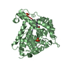
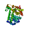
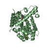
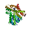
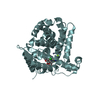
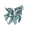
| Title | Crystal structure of PDE4D catalytic domain complexed with compound 17a | ||||||||||||
 Components Components | Isoform 3 of cAMP-specific 3',5'-cyclic phosphodiesterase 4D | ||||||||||||
 Keywords Keywords | HYDROLASE / PDE4 inhibitor | ||||||||||||
| Function / homology |  Function and homology information Function and homology informationsignaling receptor regulator activity / negative regulation of relaxation of cardiac muscle / negative regulation of heart contraction / 3',5'-cyclic-AMP phosphodiesterase / negative regulation of adenylate cyclase-activating G protein-coupled receptor signaling pathway / positive regulation of interleukin-5 production / establishment of endothelial barrier / regulation of cardiac muscle cell contraction / heterocyclic compound binding / beta-2 adrenergic receptor binding ...signaling receptor regulator activity / negative regulation of relaxation of cardiac muscle / negative regulation of heart contraction / 3',5'-cyclic-AMP phosphodiesterase / negative regulation of adenylate cyclase-activating G protein-coupled receptor signaling pathway / positive regulation of interleukin-5 production / establishment of endothelial barrier / regulation of cardiac muscle cell contraction / heterocyclic compound binding / beta-2 adrenergic receptor binding / regulation of calcium ion transmembrane transport via high voltage-gated calcium channel / voltage-gated calcium channel complex / adrenergic receptor signaling pathway / cAMP catabolic process / regulation of cell communication by electrical coupling involved in cardiac conduction / 3',5'-cyclic-nucleotide phosphodiesterase activity / 3',5'-cyclic-GMP phosphodiesterase activity / 3',5'-cyclic-AMP phosphodiesterase activity / DARPP-32 events / : / positive regulation of heart rate / cAMP binding / regulation of release of sequestered calcium ion into cytosol by sarcoplasmic reticulum / cellular response to epinephrine stimulus / calcium channel complex / positive regulation of interleukin-2 production / regulation of heart rate / cellular response to cAMP / Turbulent (oscillatory, disturbed) flow shear stress activates signaling by PIEZO1 and integrins in endothelial cells / calcium channel regulator activity / positive regulation of type II interferon production / T cell receptor signaling pathway / ATPase binding / scaffold protein binding / nuclear membrane / G alpha (s) signalling events / transmembrane transporter binding / cilium / apical plasma membrane / centrosome / enzyme binding / nucleoplasm / metal ion binding / membrane / plasma membrane / cytosol Similarity search - Function | ||||||||||||
| Biological species |  Homo sapiens (human) Homo sapiens (human) | ||||||||||||
| Method |  X-RAY DIFFRACTION / MOLECULAR REPLACEMENT / Resolution: 2.10001966681 Å X-RAY DIFFRACTION / MOLECULAR REPLACEMENT / Resolution: 2.10001966681 Å | ||||||||||||
 Authors Authors | Huang, Y.-Y. / He, X. / Luo, H.-B. | ||||||||||||
| Funding support |  China, 3items China, 3items
| ||||||||||||
 Citation Citation | Journal: J.Med.Chem. / Year: 2021 Title: Mangostanin Derivatives as Novel and Orally Active Phosphodiesterase 4 Inhibitors for the Treatment of Idiopathic Pulmonary Fibrosis with Improved Safety. Authors: Huang, Y.Y. / Deng, J. / Tian, Y.J. / Liang, J. / Xie, X. / Huang, Y. / Zhu, J. / Zhu, Z. / Zhou, Q. / He, X. / Luo, H.B. | ||||||||||||
| History |
|
- Structure visualization
| Structure viewer | Molecule: MolmilJmol/JSmol |
|---|
- Downloads & links
-Download
| PDBx/mmCIF format | 7f2k.cif.gz | 157.8 KB | Display | PDBx/mmCIF format |
|---|---|---|---|---|
| PDB format | pdb7f2k.ent.gz | 117.4 KB | Display | PDB format |
| PDBx/mmJSON format | 7f2k.json.gz | Tree view | PDBx/mmJSON format | |
| Others |  Other downloads Other downloads |
-Validation report
| Summary document | 7f2k_validation.pdf.gz | 1.1 MB | Display | wwPDB validaton report |
|---|---|---|---|---|
| Full document | 7f2k_full_validation.pdf.gz | 1.1 MB | Display | |
| Data in XML | 7f2k_validation.xml.gz | 26.8 KB | Display | |
| Data in CIF | 7f2k_validation.cif.gz | 38.7 KB | Display | |
| Arichive directory | https://data.pdbj.org/pub/pdb/validation_reports/f2/7f2kftp://data.pdbj.org/pub/pdb/validation_reports/f2/7f2k | HTTPS FTP |
-Related structure data
| Related structure data | 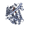 7f2lC 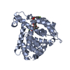 7f2mC  5wqaS S: Starting model for refinement C: citing same article ( |
|---|---|
| Similar structure data |


-Links
 PDBj
PDBj


- Assembly
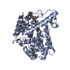
| Deposited unit | 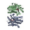
| ||||||||||||
|---|---|---|---|---|---|---|---|---|---|---|---|---|---|
| 1 | 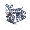
| ||||||||||||
| 2 | 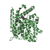
| ||||||||||||
| Unit cell |
|
-Components
| #1: Protein | Mass: 57859.504 Da / Num. of mol.: 2 Source method: isolated from a genetically manipulated source Source: (gene. exp.) Homo sapiens (human) / Gene: PDE4D, DPDE3 / Production host:  References: UniProt: Q08499, 3',5'-cyclic-AMP phosphodiesterase #2: Chemical |   Mass: 65.409 Da / Num. of mol.: 2 / Source method: obtained synthetically / Formula: Zn Mass: 65.409 Da / Num. of mol.: 2 / Source method: obtained synthetically / Formula: Zn#3: Chemical |   Mass: 24.305 Da / Num. of mol.: 2 / Source method: obtained synthetically / Formula: Mg Mass: 24.305 Da / Num. of mol.: 2 / Source method: obtained synthetically / Formula: Mg#4: Chemical |   Mass: 492.517 Da / Num. of mol.: 2 / Source method: obtained synthetically / Formula: C28H28O8 / Feature type: SUBJECT OF INVESTIGATION Mass: 492.517 Da / Num. of mol.: 2 / Source method: obtained synthetically / Formula: C28H28O8 / Feature type: SUBJECT OF INVESTIGATION#5: Water | ChemComp-HOH / |  Mass: 18.015 Da / Num. of mol.: 288 / Source method: isolated from a natural source / Formula: H2O Mass: 18.015 Da / Num. of mol.: 288 / Source method: isolated from a natural source / Formula: H2OHas ligand of interest | Y | |
|---|
-Experimental details
-Experiment
| Experiment | Method: X-RAY DIFFRACTION / Number of used crystals: 1 |
|---|
- Sample preparation
| Crystal | Density Matthews: 1.65 Å3/Da / Density % sol: 25.63 % |
|---|---|
| Crystal grow | Temperature: 277 K / Method: vapor diffusion Details: 0.1 M HEPES (pH 7.4), 0.1 M MgCl2, 15% PEG3350, 10% isopropanol, and 25% ethylene glycol |
-Data collection
| Diffraction | Mean temperature: 100 K / Serial crystal experiment: N |
|---|---|
| Diffraction source | Source: ROTATING ANODE / Type: RIGAKU / Wavelength: 1.5418 Å |
| Detector | Type: RIGAKU HyPix-3000 / Detector: PIXEL / Date: Mar 22, 2021 |
| Radiation | Protocol: SINGLE WAVELENGTH / Monochromatic (M) / Laue (L): M / Scattering type: x-ray |
| Radiation wavelength | Wavelength: 1.5418 Å / Relative weight: 1 |
| Reflection | Resolution: 2.1→23.58 Å / Num. obs: 45574 / % possible obs: 99.8 % / Redundancy: 4.3 % / Biso Wilson estimate: 21.3256254028 Å2 / Rmerge(I) obs: 0.075 / Net I/σ(I): 15.3 |
| Reflection shell | Resolution: 2.1→2.2 Å / Redundancy: 2.8 % / Rmerge(I) obs: 0.311 / Mean I/σ(I) obs: 2.8 / Num. unique obs: 4490 / % possible all: 99.6 |
- Processing
| Software |
| |||||||||||||||||||||||||||||||||||||||||||||||||||||||||||||||||||||||||||||||||||||||||||||||||||||||||||||||||||||||
|---|---|---|---|---|---|---|---|---|---|---|---|---|---|---|---|---|---|---|---|---|---|---|---|---|---|---|---|---|---|---|---|---|---|---|---|---|---|---|---|---|---|---|---|---|---|---|---|---|---|---|---|---|---|---|---|---|---|---|---|---|---|---|---|---|---|---|---|---|---|---|---|---|---|---|---|---|---|---|---|---|---|---|---|---|---|---|---|---|---|---|---|---|---|---|---|---|---|---|---|---|---|---|---|---|---|---|---|---|---|---|---|---|---|---|---|---|---|---|---|---|
| Refinement | Method to determine structure: MOLECULAR REPLACEMENT Starting model: 5WQA Resolution: 2.10001966681→23.5783341262 Å / SU ML: 0.253969836619 / Cross valid method: NONE / σ(F): 1.35988904769 / Phase error: 24.0687303043 Stereochemistry target values: GeoStd + Monomer Library + CDL v1.2
| |||||||||||||||||||||||||||||||||||||||||||||||||||||||||||||||||||||||||||||||||||||||||||||||||||||||||||||||||||||||
| Solvent computation | Shrinkage radii: 0.9 Å / VDW probe radii: 1.11 Å / Solvent model: FLAT BULK SOLVENT MODEL | |||||||||||||||||||||||||||||||||||||||||||||||||||||||||||||||||||||||||||||||||||||||||||||||||||||||||||||||||||||||
| Displacement parameters | Biso mean: 27.2593589744 Å2 | |||||||||||||||||||||||||||||||||||||||||||||||||||||||||||||||||||||||||||||||||||||||||||||||||||||||||||||||||||||||
| Refinement step | Cycle: LAST / Resolution: 2.10001966681→23.5783341262 Å
| |||||||||||||||||||||||||||||||||||||||||||||||||||||||||||||||||||||||||||||||||||||||||||||||||||||||||||||||||||||||
| Refine LS restraints |
| |||||||||||||||||||||||||||||||||||||||||||||||||||||||||||||||||||||||||||||||||||||||||||||||||||||||||||||||||||||||
| LS refinement shell |
|
