 Movie
Movie Controller

[English] 日本語
 Yorodumi
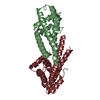
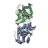
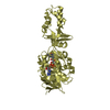
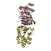
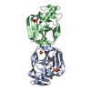
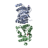
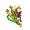
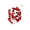
- PDB-7ev8: Structure of Human Parainfluenza Virus 3 Unassembled Nucleoprotei... -
+ Open data

- Basic information
| Entry | Database: PDB / ID: 7ev8 | ||||||
|---|---|---|---|---|---|---|---|
| Title | Structure of Human Parainfluenza Virus 3 Unassembled Nucleoprotein in Complex with its viral chaperone | ||||||
 Components Components |
| ||||||
 Keywords Keywords | VIRAL PROTEIN / Complex / Inhibitor / HPIV3 / Nucleoprotein | ||||||
| Function / homology |  Function and homology information Function and homology informationhelical viral capsid / viral genome replication / viral nucleocapsid / host cell cytoplasm / ribonucleoprotein complex / RNA-directed RNA polymerase activity / DNA-templated transcription / structural molecule activity / RNA binding Similarity search - Function | ||||||
| Biological species |  Human parainfluenza 3 virusHuman respirovirus 3 Human parainfluenza 3 virusHuman respirovirus 3 | ||||||
| Method |  X-RAY DIFFRACTION / SYNCHROTRON / MOLECULAR REPLACEMENT / Resolution: 3.234 Å X-RAY DIFFRACTION / SYNCHROTRON / MOLECULAR REPLACEMENT / Resolution: 3.234 Å | ||||||
 Authors Authors | Dong, X.F. / Chen, Z. | ||||||
| Funding support |  China, 1items China, 1items
| ||||||
 Citation Citation | Journal: J.Virol. / Year: 2022 Title: Structural Basis of Human Parainfluenza Virus 3 Unassembled Nucleoprotein in Complex with Its Viral Chaperone. Authors: Dong, X. / Wang, X. / Xie, M. / Wu, W. / Chen, Z. | ||||||
| History |
|
- Structure visualization
| Structure viewer | Molecule: MolmilJmol/JSmol |
|---|
- Downloads & links
-Download
| PDBx/mmCIF format | 7ev8.cif.gz | 87.2 KB | Display | PDBx/mmCIF format |
|---|---|---|---|---|
| PDB format | pdb7ev8.ent.gz | 60.3 KB | Display | PDB format |
| PDBx/mmJSON format | 7ev8.json.gz | Tree view | PDBx/mmJSON format | |
| Others |  Other downloads Other downloads |
-Validation report
| Summary document | 7ev8_validation.pdf.gz | 431.1 KB | Display | wwPDB validaton report |
|---|---|---|---|---|
| Full document | 7ev8_full_validation.pdf.gz | 433 KB | Display | |
| Data in XML | 7ev8_validation.xml.gz | 14.2 KB | Display | |
| Data in CIF | 7ev8_validation.cif.gz | 19.1 KB | Display | |
| Arichive directory | https://data.pdbj.org/pub/pdb/validation_reports/ev/7ev8ftp://data.pdbj.org/pub/pdb/validation_reports/ev/7ev8 | HTTPS FTP |
-Related structure data
| Related structure data | 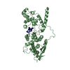 6h5qS S: Starting model for refinement |
|---|---|
| Similar structure data |


-Links
 PDBj
PDBj

- Assembly
| Deposited unit | 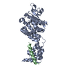
| ||||||||
|---|---|---|---|---|---|---|---|---|---|
| 1 |
| ||||||||
| Unit cell |
|
-Components
| #1: Protein | Mass: 38831.461 Da / Num. of mol.: 1 Source method: isolated from a genetically manipulated source Source: (gene. exp.) Human parainfluenza 3 virus (strain Wash/47885/57)Strain: Wash/47885/57 / Gene: N, NP / Production host:  |
|---|---|
| #2: Protein/peptide | Mass: 4879.281 Da / Num. of mol.: 1 Source method: isolated from a genetically manipulated source Source: (gene. exp.) Human respirovirus 3 / Production host: |
| #3: Water | ChemComp-HOH /  Mass: 18.015 Da / Num. of mol.: 54 / Source method: isolated from a natural source / Formula: H2O Mass: 18.015 Da / Num. of mol.: 54 / Source method: isolated from a natural source / Formula: H2O |
-Experimental details
-Experiment
| Experiment | Method: X-RAY DIFFRACTION / Number of used crystals: 1 |
|---|
- Sample preparation
| Crystal | Density Matthews: 2.81 Å3/Da / Density % sol: 56.18 % |
|---|---|
| Crystal grow | Temperature: 277.15 K / Method: vapor diffusion, sitting drop / Details: PEG 6000, Cacodylate, Nickel chloride |
-Data collection
| Diffraction | Mean temperature: 80 K / Serial crystal experiment: N | |||||||||||||||||||||||||||||||||||||||||||||||||||||||||||||||||||||||||||||||||||||||||||||||||||||||||||||||||||||||||||||||||||||||||||||||||||||||||||||||||||||||||||||||||||||||||||||
|---|---|---|---|---|---|---|---|---|---|---|---|---|---|---|---|---|---|---|---|---|---|---|---|---|---|---|---|---|---|---|---|---|---|---|---|---|---|---|---|---|---|---|---|---|---|---|---|---|---|---|---|---|---|---|---|---|---|---|---|---|---|---|---|---|---|---|---|---|---|---|---|---|---|---|---|---|---|---|---|---|---|---|---|---|---|---|---|---|---|---|---|---|---|---|---|---|---|---|---|---|---|---|---|---|---|---|---|---|---|---|---|---|---|---|---|---|---|---|---|---|---|---|---|---|---|---|---|---|---|---|---|---|---|---|---|---|---|---|---|---|---|---|---|---|---|---|---|---|---|---|---|---|---|---|---|---|---|---|---|---|---|---|---|---|---|---|---|---|---|---|---|---|---|---|---|---|---|---|---|---|---|---|---|---|---|---|---|---|---|---|
| Diffraction source | Source: SYNCHROTRON / Site: SSRF / Beamline: BL17U1 / Wavelength: 0.979 Å | |||||||||||||||||||||||||||||||||||||||||||||||||||||||||||||||||||||||||||||||||||||||||||||||||||||||||||||||||||||||||||||||||||||||||||||||||||||||||||||||||||||||||||||||||||||||||||||
| Detector | Type: DECTRIS EIGER X 16M / Detector: PIXEL / Date: Jun 22, 2020 | |||||||||||||||||||||||||||||||||||||||||||||||||||||||||||||||||||||||||||||||||||||||||||||||||||||||||||||||||||||||||||||||||||||||||||||||||||||||||||||||||||||||||||||||||||||||||||||
| Radiation | Protocol: SINGLE WAVELENGTH / Monochromatic (M) / Laue (L): M / Scattering type: x-ray | |||||||||||||||||||||||||||||||||||||||||||||||||||||||||||||||||||||||||||||||||||||||||||||||||||||||||||||||||||||||||||||||||||||||||||||||||||||||||||||||||||||||||||||||||||||||||||||
| Radiation wavelength | Wavelength: 0.979 Å / Relative weight: 1 | |||||||||||||||||||||||||||||||||||||||||||||||||||||||||||||||||||||||||||||||||||||||||||||||||||||||||||||||||||||||||||||||||||||||||||||||||||||||||||||||||||||||||||||||||||||||||||||
| Reflection | Resolution: 3.23→50 Å / Num. obs: 8248 / % possible obs: 99.8 % / Redundancy: 6.8 % / Rmerge(I) obs: 0.098 / Rpim(I) all: 0.04 / Rrim(I) all: 0.106 / Χ2: 0.903 / Net I/σ(I): 9.1 / Num. measured all: 56267 | |||||||||||||||||||||||||||||||||||||||||||||||||||||||||||||||||||||||||||||||||||||||||||||||||||||||||||||||||||||||||||||||||||||||||||||||||||||||||||||||||||||||||||||||||||||||||||||
| Reflection shell | Diffraction-ID: 1
|
- Processing
| Software |
| |||||||||||||||||||||||||||||||||||||||||||||||||||||||||||||||||||||||||||||||||||||||||||||||||||||||||||||||||||||||||||||||||||||||||||||||||||||||||||
|---|---|---|---|---|---|---|---|---|---|---|---|---|---|---|---|---|---|---|---|---|---|---|---|---|---|---|---|---|---|---|---|---|---|---|---|---|---|---|---|---|---|---|---|---|---|---|---|---|---|---|---|---|---|---|---|---|---|---|---|---|---|---|---|---|---|---|---|---|---|---|---|---|---|---|---|---|---|---|---|---|---|---|---|---|---|---|---|---|---|---|---|---|---|---|---|---|---|---|---|---|---|---|---|---|---|---|---|---|---|---|---|---|---|---|---|---|---|---|---|---|---|---|---|---|---|---|---|---|---|---|---|---|---|---|---|---|---|---|---|---|---|---|---|---|---|---|---|---|---|---|---|---|---|---|---|---|
| Refinement | Method to determine structure: MOLECULAR REPLACEMENT Starting model: 6H5Q Resolution: 3.234→49.178 Å / Cor.coef. Fo:Fc: 0.899 / Cor.coef. Fo:Fc free: 0.842 / WRfactor Rfree: 0.243 / WRfactor Rwork: 0.205 / SU B: 14.998 / SU ML: 0.27 / Average fsc free: 0.9682 / Average fsc work: 0.9757 / Cross valid method: THROUGHOUT / ESU R Free: 0.1 Details: Hydrogens have been added in their riding positions
| |||||||||||||||||||||||||||||||||||||||||||||||||||||||||||||||||||||||||||||||||||||||||||||||||||||||||||||||||||||||||||||||||||||||||||||||||||||||||||
| Solvent computation | Ion probe radii: 0.8 Å / Shrinkage radii: 0.8 Å / VDW probe radii: 1.2 Å / Solvent model: MASK BULK SOLVENT | |||||||||||||||||||||||||||||||||||||||||||||||||||||||||||||||||||||||||||||||||||||||||||||||||||||||||||||||||||||||||||||||||||||||||||||||||||||||||||
| Displacement parameters | Biso mean: 45.392 Å2
| |||||||||||||||||||||||||||||||||||||||||||||||||||||||||||||||||||||||||||||||||||||||||||||||||||||||||||||||||||||||||||||||||||||||||||||||||||||||||||
| Refinement step | Cycle: LAST / Resolution: 3.234→49.178 Å
| |||||||||||||||||||||||||||||||||||||||||||||||||||||||||||||||||||||||||||||||||||||||||||||||||||||||||||||||||||||||||||||||||||||||||||||||||||||||||||
| Refine LS restraints |
| |||||||||||||||||||||||||||||||||||||||||||||||||||||||||||||||||||||||||||||||||||||||||||||||||||||||||||||||||||||||||||||||||||||||||||||||||||||||||||
| LS refinement shell |
|
