 Movie
Movie Controller

[English] 日本語
 Yorodumi
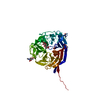
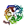
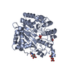
- PDB-7esk: Crystal structure of a L-rhamnose-alpha-1,4-D-glucuronate lyase f... -
+ Open data

- Basic information
| Entry | Database: PDB / ID: 7esk | ||||||
|---|---|---|---|---|---|---|---|
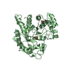
| Title | Crystal structure of a L-rhamnose-alpha-1,4-D-glucuronate lyase from Fusarium oxysporum 12S, Ligand free form | ||||||
 Components Components | L-Rhamnose-alpha-1,4-D-glucuronate lyase | ||||||
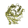
 Keywords Keywords | LYASE / seven-bladed beta-propeller | ||||||
| Biological species |   Fusarium oxysporum (fungus) Fusarium oxysporum (fungus) | ||||||
| Method |  X-RAY DIFFRACTION / SYNCHROTRON / SAD / Resolution: 1.05 Å X-RAY DIFFRACTION / SYNCHROTRON / SAD / Resolution: 1.05 Å | ||||||
 Authors Authors | Kondo, T. / Arakawa, T. / Fushinobu, S. / Sakamoto, T. | ||||||
 Citation Citation | Journal: J.Biol.Chem. / Year: 2021 Title: Structural and functional analysis of gum arabic l-rhamnose-alpha-1,4-d-glucuronate lyase establishes a novel polysaccharide lyase family. Authors: Kondo, T. / Kichijo, M. / Maruta, A. / Nakaya, M. / Takenaka, S. / Arakawa, T. / Fushinobu, S. / Sakamoto, T. | ||||||
| History |
|
- Structure visualization
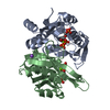
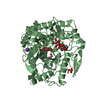
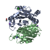
| Structure viewer | Molecule:  MolmilJmol/JSmol MolmilJmol/JSmol |
|---|
- Downloads & links
-Download
| PDBx/mmCIF format | 7esk.cif.gz | 116.1 KB | Display | PDBx/mmCIF format |
|---|---|---|---|---|
| PDB format | pdb7esk.ent.gz | 85.6 KB | Display | PDB format |
| PDBx/mmJSON format | 7esk.json.gz | Tree view | PDBx/mmJSON format | |
| Others |  Other downloads Other downloads |
-Validation report
| Arichive directory | https://data.pdbj.org/pub/pdb/validation_reports/es/7eskftp://data.pdbj.org/pub/pdb/validation_reports/es/7esk | HTTPS FTP |
|---|
-Related structure data


-Links
 PDBj
PDBj- Assembly
| Deposited unit | 
| ||||||||
|---|---|---|---|---|---|---|---|---|---|
| 1 |
| ||||||||
| Unit cell |
|
-Components
| #1: Protein | Mass: 50085.199 Da / Num. of mol.: 1 Source method: isolated from a genetically manipulated source Source: (gene. exp.) Fusarium oxysporum (fungus) / Strain: 12S / Gene: Forham1 / Plasmid: pPICZ-alpha-A / Production host: Komagataella pastoris (fungus) / Strain (production host): X-33References: Lyases; Carbon-oxygen lyases; Acting on polysaccharides |
|---|---|
| #2: Polysaccharide | alpha-D-mannopyranose-(1-3)-beta-D-mannopyranose-(1-4)-2-acetamido-2-deoxy-beta-D-glucopyranose-(1- ...alpha-D-mannopyranose-(1-3)-beta-D-mannopyranose-(1-4)-2-acetamido-2-deoxy-beta-D-glucopyranose-(1-4)-2-acetamido-2-deoxy-beta-D-glucopyranose Source method: isolated from a genetically manipulated source |
| #3: Chemical | ChemComp-CA /   Mass: 40.078 Da / Num. of mol.: 1 / Source method: isolated from a natural source / Formula: Ca / Feature type: SUBJECT OF INVESTIGATION Mass: 40.078 Da / Num. of mol.: 1 / Source method: isolated from a natural source / Formula: Ca / Feature type: SUBJECT OF INVESTIGATION |
| #4: Chemical | ChemComp-NA /   Mass: 22.990 Da / Num. of mol.: 1 / Source method: obtained synthetically / Formula: Na / Feature type: SUBJECT OF INVESTIGATION Mass: 22.990 Da / Num. of mol.: 1 / Source method: obtained synthetically / Formula: Na / Feature type: SUBJECT OF INVESTIGATION |
| #5: Water | ChemComp-HOH /  Mass: 18.015 Da / Num. of mol.: 597 / Source method: isolated from a natural source / Formula: H2O Mass: 18.015 Da / Num. of mol.: 597 / Source method: isolated from a natural source / Formula: H2O |
| Has ligand of interest | Y |
| Has protein modification | Y |
| Sequence details | The amino acid sequence of FoRham1 has been registered in GenBank, DDBj and EMBL. Its accession ...The amino acid sequence of FoRham1 has been registered in GenBank, DDBj and EMBL. Its accession number is LC617219. |
-Experimental details
-Experiment
| Experiment | Method: X-RAY DIFFRACTION / Number of used crystals: 1 |
|---|
- Sample preparation
| Crystal | Density Matthews: 2.03 Å3/Da / Density % sol: 39.29 % |
|---|---|
| Crystal grow | Temperature: 293.15 K / Method: vapor diffusion, hanging drop / pH: 8.5 Details: 30 % (v/v) PEG 1500, 0.1 M BICINE-NaOH (pH 8.5), 10 % (v/v) 2-propanol, Crystal was soaked into 20 % (v/v) glycerol at 298 K for 1 min |
-Data collection
| Diffraction | Mean temperature: 100 K / Serial crystal experiment: N |
|---|---|
| Diffraction source | Source: SYNCHROTRON / Site: Photon Factory  / Beamline: BL-5A / Wavelength: 1 Å / Beamline: BL-5A / Wavelength: 1 Å |
| Detector | Type: DECTRIS PILATUS3 S 6M / Detector: PIXEL / Date: Mar 2, 2019 |
| Radiation | Monochromator: Numerical link type Si(111) double crystal / Protocol: SINGLE WAVELENGTH / Monochromatic (M) / Laue (L): M / Scattering type: x-ray |
| Radiation wavelength | Wavelength: 1 Å / Relative weight: 1 |
| Reflection | Resolution: 1.05→50 Å / Num. obs: 190044 / % possible obs: 94.2 % / Redundancy: 6.2 % / CC1/2: 0.999 / Net I/σ(I): 39.4 |
| Reflection shell | Resolution: 1.05→1.07 Å / Num. unique obs: 9391 / CC1/2: 0.931 |
- Processing
| Software |
| ||||||||||||||||||||||||||||||||||||||||||||||||||||||||||||
|---|---|---|---|---|---|---|---|---|---|---|---|---|---|---|---|---|---|---|---|---|---|---|---|---|---|---|---|---|---|---|---|---|---|---|---|---|---|---|---|---|---|---|---|---|---|---|---|---|---|---|---|---|---|---|---|---|---|---|---|---|---|
| Refinement | Method to determine structure: SAD / Resolution: 1.05→31.3 Å / Cor.coef. Fo:Fc: 0.98 / Cor.coef. Fo:Fc free: 0.973 / SU B: 0.389 / SU ML: 0.019 / Cross valid method: THROUGHOUT / σ(F): 0 / ESU R: 0.025 / ESU R Free: 0.027 / Stereochemistry target values: MAXIMUM LIKELIHOOD Details: HYDROGENS HAVE BEEN ADDED IN THE RIDING POSITIONS U VALUES : REFINED INDIVIDUALLY
| ||||||||||||||||||||||||||||||||||||||||||||||||||||||||||||
| Solvent computation | Ion probe radii: 0.8 Å / Shrinkage radii: 0.8 Å / VDW probe radii: 1.2 Å / Solvent model: BABINET MODEL WITH MASK | ||||||||||||||||||||||||||||||||||||||||||||||||||||||||||||
| Displacement parameters | Biso max: 54.71 Å2 / Biso mean: 16.018 Å2 / Biso min: 9.25 Å2
| ||||||||||||||||||||||||||||||||||||||||||||||||||||||||||||
| Refinement step | Cycle: final / Resolution: 1.05→31.3 Å
| ||||||||||||||||||||||||||||||||||||||||||||||||||||||||||||
| Refine LS restraints |
| ||||||||||||||||||||||||||||||||||||||||||||||||||||||||||||
| LS refinement shell | Resolution: 1.05→1.077 Å / Rfactor Rfree error: 0 / Total num. of bins used: 20
|
