 Movie
Movie Controller


+ Open data

- Basic information
| Entry | Database: PDB / ID: 7eps | ||||||||||||
|---|---|---|---|---|---|---|---|---|---|---|---|---|---|
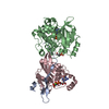
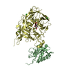
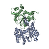
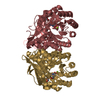
| Title | Partial Consensus L-threonine 3-dehydrogenase (E-change) | ||||||||||||
 Components Components | L-threonine 3-dehydrogenase | ||||||||||||
 Keywords Keywords | OXIDOREDUCTASE / L-threonine 3-dehydrogenase / artificial protein | ||||||||||||
| Function / homology | NICOTINAMIDE-ADENINE-DINUCLEOTIDE / THREONINE Function and homology information Function and homology information | ||||||||||||
| Biological species | synthetic construct (others) | ||||||||||||
| Method |  X-RAY DIFFRACTION / SYNCHROTRON / MOLECULAR REPLACEMENT / Resolution: 2.102 Å X-RAY DIFFRACTION / SYNCHROTRON / MOLECULAR REPLACEMENT / Resolution: 2.102 Å | ||||||||||||
 Authors Authors | Kozuka, K. / Nakano, S. / Asano, Y. / Ito, S. | ||||||||||||
| Funding support |  Japan, 3items Japan, 3items
| ||||||||||||
 Citation Citation | Journal: Biochemistry / Year: 2021 Title: Partial Consensus Design and Enhancement of Protein Function by Secondary-Structure-Guided Consensus Mutations. Authors: Kozuka, K. / Nakano, S. / Asano, Y. / Ito, S. | ||||||||||||
| History |
|
- Structure visualization
| Structure viewer | Molecule: MolmilJmol/JSmol |
|---|
- Downloads & links
-Download
| PDBx/mmCIF format | 7eps.cif.gz | 271 KB | Display | PDBx/mmCIF format |
|---|---|---|---|---|
| PDB format | pdb7eps.ent.gz | 210 KB | Display | PDB format |
| PDBx/mmJSON format | 7eps.json.gz | Tree view | PDBx/mmJSON format | |
| Others |  Other downloads Other downloads |
-Validation report
| Arichive directory | https://data.pdbj.org/pub/pdb/validation_reports/ep/7epsftp://data.pdbj.org/pub/pdb/validation_reports/ep/7eps | HTTPS FTP |
|---|
-Related structure data
| Related structure data |  7eprC  3wmxS S: Starting model for refinement C: citing same article ( |
|---|---|
| Similar structure data |
-Links
 PDBj
PDBj- Assembly
| Deposited unit | 
| ||||||||||||||||||||||||||||||||||||||||||||||||||||||||||||||||||||||||||||||||||||||||||||||||||||||||||||||||||||||
|---|---|---|---|---|---|---|---|---|---|---|---|---|---|---|---|---|---|---|---|---|---|---|---|---|---|---|---|---|---|---|---|---|---|---|---|---|---|---|---|---|---|---|---|---|---|---|---|---|---|---|---|---|---|---|---|---|---|---|---|---|---|---|---|---|---|---|---|---|---|---|---|---|---|---|---|---|---|---|---|---|---|---|---|---|---|---|---|---|---|---|---|---|---|---|---|---|---|---|---|---|---|---|---|---|---|---|---|---|---|---|---|---|---|---|---|---|---|---|---|
| 1 | 
| ||||||||||||||||||||||||||||||||||||||||||||||||||||||||||||||||||||||||||||||||||||||||||||||||||||||||||||||||||||||
| 2 | 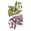
| ||||||||||||||||||||||||||||||||||||||||||||||||||||||||||||||||||||||||||||||||||||||||||||||||||||||||||||||||||||||
| Unit cell |
| ||||||||||||||||||||||||||||||||||||||||||||||||||||||||||||||||||||||||||||||||||||||||||||||||||||||||||||||||||||||
| Noncrystallographic symmetry (NCS) | NCS domain:
NCS domain segments: Beg auth comp-ID: PRO / Beg label comp-ID: PRO / End auth comp-ID: THR / End label comp-ID: THR / Auth seq-ID: 6 - 315 / Label seq-ID: 26 - 335
NCS ensembles :
|
-Components
| #1: Protein | Mass: 36819.676 Da / Num. of mol.: 4 Source method: isolated from a genetically manipulated source Source: (gene. exp.) synthetic construct (others) / Production host:  #2: Chemical | ChemComp-NAD /   Mass: 663.425 Da / Num. of mol.: 4 / Source method: obtained synthetically / Formula: C21H27N7O14P2 / Feature type: SUBJECT OF INVESTIGATION / Comment: NAD*YM Mass: 663.425 Da / Num. of mol.: 4 / Source method: obtained synthetically / Formula: C21H27N7O14P2 / Feature type: SUBJECT OF INVESTIGATION / Comment: NAD*YM#3: Chemical | ChemComp-THR /   Type: L-peptide linking / Mass: 119.119 Da / Num. of mol.: 4 / Source method: obtained synthetically / Formula: C4H9NO3 / Feature type: SUBJECT OF INVESTIGATION Type: L-peptide linking / Mass: 119.119 Da / Num. of mol.: 4 / Source method: obtained synthetically / Formula: C4H9NO3 / Feature type: SUBJECT OF INVESTIGATION#4: Chemical |   Mass: 354.436 Da / Num. of mol.: 2 / Source method: obtained synthetically / Formula: C16H34O8 / Feature type: SUBJECT OF INVESTIGATION / Comment: precipitant*YM Mass: 354.436 Da / Num. of mol.: 2 / Source method: obtained synthetically / Formula: C16H34O8 / Feature type: SUBJECT OF INVESTIGATION / Comment: precipitant*YM#5: Water | ChemComp-HOH / |  Mass: 18.015 Da / Num. of mol.: 574 / Source method: isolated from a natural source / Formula: H2O Mass: 18.015 Da / Num. of mol.: 574 / Source method: isolated from a natural source / Formula: H2OHas ligand of interest | Y | |
|---|
-Experimental details
-Experiment
| Experiment | Method: X-RAY DIFFRACTION / Number of used crystals: 1 |
|---|
- Sample preparation
| Crystal | Density Matthews: 3.75 Å3/Da / Density % sol: 67.18 % |
|---|---|
| Crystal grow | Temperature: 295 K / Method: vapor diffusion, sitting drop / Details: 20% [w/v] PEG 3350, 0.2M magnesium acetate |
-Data collection
| Diffraction | Mean temperature: 100 K / Serial crystal experiment: N |
|---|---|
| Diffraction source | Source: SYNCHROTRON / Site: Photon Factory / Beamline: BL-5A / Wavelength: 1 Å |
| Detector | Type: DECTRIS PILATUS3 6M / Detector: PIXEL / Date: Mar 5, 2020 |
| Radiation | Protocol: SINGLE WAVELENGTH / Monochromatic (M) / Laue (L): M / Scattering type: x-ray |
| Radiation wavelength | Wavelength: 1 Å / Relative weight: 1 |
| Reflection | Resolution: 2.1→49.4 Å / Num. obs: 878278 / % possible obs: 100 % / Redundancy: 7 % / CC1/2: 1 / Rmerge(I) obs: 0.087 / Net I/σ(I): 15.5 |
| Reflection shell | Resolution: 2.1→2.22 Å / Rmerge(I) obs: 0.57 / Num. unique obs: 18253 / CC1/2: 0.895 |
- Processing
| Software |
| ||||||||||||||||||||||||||||||||||||||||||||||||||||||||||||||||||||||||||||||||||||||||||||||||||||||||||||||||||||||||||||||||||||||||||||||||||||||||||||||||||||||||||||||||||||||
|---|---|---|---|---|---|---|---|---|---|---|---|---|---|---|---|---|---|---|---|---|---|---|---|---|---|---|---|---|---|---|---|---|---|---|---|---|---|---|---|---|---|---|---|---|---|---|---|---|---|---|---|---|---|---|---|---|---|---|---|---|---|---|---|---|---|---|---|---|---|---|---|---|---|---|---|---|---|---|---|---|---|---|---|---|---|---|---|---|---|---|---|---|---|---|---|---|---|---|---|---|---|---|---|---|---|---|---|---|---|---|---|---|---|---|---|---|---|---|---|---|---|---|---|---|---|---|---|---|---|---|---|---|---|---|---|---|---|---|---|---|---|---|---|---|---|---|---|---|---|---|---|---|---|---|---|---|---|---|---|---|---|---|---|---|---|---|---|---|---|---|---|---|---|---|---|---|---|---|---|---|---|---|---|
| Refinement | Method to determine structure: MOLECULAR REPLACEMENT Starting model: 3WMX Resolution: 2.102→49.4 Å / Cor.coef. Fo:Fc: 0.94 / Cor.coef. Fo:Fc free: 0.923 / SU B: 3.947 / SU ML: 0.103 / Cross valid method: FREE R-VALUE / ESU R: 0.161 / ESU R Free: 0.148 Details: Hydrogens have been added in their riding positions
| ||||||||||||||||||||||||||||||||||||||||||||||||||||||||||||||||||||||||||||||||||||||||||||||||||||||||||||||||||||||||||||||||||||||||||||||||||||||||||||||||||||||||||||||||||||||
| Solvent computation | Ion probe radii: 0.8 Å / Shrinkage radii: 0.8 Å / VDW probe radii: 1.2 Å / Solvent model: MASK BULK SOLVENT | ||||||||||||||||||||||||||||||||||||||||||||||||||||||||||||||||||||||||||||||||||||||||||||||||||||||||||||||||||||||||||||||||||||||||||||||||||||||||||||||||||||||||||||||||||||||
| Displacement parameters | Biso mean: 31.893 Å2
| ||||||||||||||||||||||||||||||||||||||||||||||||||||||||||||||||||||||||||||||||||||||||||||||||||||||||||||||||||||||||||||||||||||||||||||||||||||||||||||||||||||||||||||||||||||||
| Refinement step | Cycle: LAST / Resolution: 2.102→49.4 Å
| ||||||||||||||||||||||||||||||||||||||||||||||||||||||||||||||||||||||||||||||||||||||||||||||||||||||||||||||||||||||||||||||||||||||||||||||||||||||||||||||||||||||||||||||||||||||
| Refine LS restraints |
|