Movie
Movie Controller

[English] 日本語
 Yorodumi
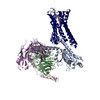
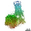
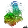
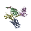
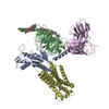
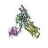
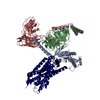
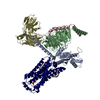
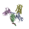
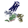
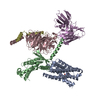
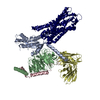
- PDB-7eo2: Cryo-EM of Sphingosine 1-phosphate receptor 1 / Gi complex bound ... -
+ Open data

- Basic information
| Entry | Database: PDB / ID: 7eo2 | ||||||
|---|---|---|---|---|---|---|---|
| Title | Cryo-EM of Sphingosine 1-phosphate receptor 1 / Gi complex bound to FTY720p | ||||||
 Components Components |
| ||||||
 Keywords Keywords | MEMBRANE PROTEIN / GPCR | ||||||
| Function / homology |  Function and homology information Function and homology informationcardiac muscle tissue growth involved in heart morphogenesis / blood vessel maturation / sphingolipid binding / sphingosine-1-phosphate receptor activity / Lysosphingolipid and LPA receptors / endothelial cell differentiation / heart trabecula morphogenesis / regulation of bone mineralization / negative regulation of stress fiber assembly / sphingosine-1-phosphate receptor signaling pathway ...cardiac muscle tissue growth involved in heart morphogenesis / blood vessel maturation / sphingolipid binding / sphingosine-1-phosphate receptor activity / Lysosphingolipid and LPA receptors / endothelial cell differentiation / heart trabecula morphogenesis / regulation of bone mineralization / negative regulation of stress fiber assembly / sphingosine-1-phosphate receptor signaling pathway / leukocyte chemotaxis / positive regulation of positive chemotaxis / regulation of bone resorption / lamellipodium assembly / transmission of nerve impulse / regulation of cell adhesion / adenylate cyclase inhibitor activity / positive regulation of protein localization to cell cortex / T cell migration / positive regulation of relaxation of smooth muscle / Adenylate cyclase inhibitory pathway / D2 dopamine receptor binding / positive regulation of smooth muscle cell proliferation / adenylate cyclase-inhibiting serotonin receptor signaling pathway / G protein-coupled serotonin receptor binding / cellular response to forskolin / regulation of mitotic spindle organization / chemokine-mediated signaling pathway / Regulation of insulin secretion / neuropeptide signaling pathway / response to prostaglandin E / brain development / positive regulation of cholesterol biosynthetic process / negative regulation of insulin secretion / G protein-coupled receptor binding / response to peptide hormone / G protein-coupled receptor activity / chemotaxis / centriolar satellite / neuron differentiation / G-protein beta/gamma-subunit complex binding / adenylate cyclase-modulating G protein-coupled receptor signaling pathway / adenylate cyclase-inhibiting G protein-coupled receptor signaling pathway / Olfactory Signaling Pathway / Activation of the phototransduction cascade / G protein-coupled acetylcholine receptor signaling pathway / G beta:gamma signalling through PLC beta / Presynaptic function of Kainate receptors / Thromboxane signalling through TP receptor / Activation of G protein gated Potassium channels / Inhibition of voltage gated Ca2+ channels via Gbeta/gamma subunits / G-protein activation / Glucagon signaling in metabolic regulation / Prostacyclin signalling through prostacyclin receptor / G beta:gamma signalling through CDC42 / Synthesis, secretion, and inactivation of Glucagon-like Peptide-1 (GLP-1) / G beta:gamma signalling through BTK / photoreceptor disc membrane / ADP signalling through P2Y purinoceptor 12 / Glucagon-type ligand receptors / GDP binding / Sensory perception of sweet, bitter, and umami (glutamate) taste / Adrenaline,noradrenaline inhibits insulin secretion / Vasopressin regulates renal water homeostasis via Aquaporins / Glucagon-like Peptide-1 (GLP1) regulates insulin secretion / G alpha (z) signalling events / cellular response to catecholamine stimulus / ADP signalling through P2Y purinoceptor 1 / ADORA2B mediated anti-inflammatory cytokines production / G beta:gamma signalling through PI3Kgamma / cell migration / adenylate cyclase-activating dopamine receptor signaling pathway / Cooperation of PDCL (PhLP1) and TRiC/CCT in G-protein beta folding / GPER1 signaling / cellular response to prostaglandin E stimulus / heterotrimeric G-protein complex / G alpha (12/13) signalling events / Inactivation, recovery and regulation of the phototransduction cascade / G-protein beta-subunit binding / extracellular vesicle / sensory perception of taste / sperm principal piece / Thrombin signalling through proteinase activated receptors (PARs) / adenylate cyclase-activating G protein-coupled receptor signaling pathway / presynapse / signaling receptor complex adaptor activity / actin cytoskeleton organization / retina development in camera-type eye / Interleukin-4 and Interleukin-13 signaling / GTPase binding / angiogenesis / fibroblast proliferation / G protein activity / midbody / Ca2+ pathway / cell cortex / High laminar flow shear stress activates signaling by PIEZO1 and PECAM1:CDH5:KDR in endothelial cells / Potential therapeutics for SARS / G alpha (i) signalling events / G alpha (s) signalling events Similarity search - Function | ||||||
| Biological species |  Homo sapiens (human) Homo sapiens (human) | ||||||
| Method | ELECTRON MICROSCOPY / single particle reconstruction / cryo EM / Resolution: 2.89 Å | ||||||
 Authors Authors | He, Y. / Xu, Z. / Ikuta, T. | ||||||
| Funding support |  China, 1items China, 1items
| ||||||
 Citation Citation | Journal: Nat Chem Biol / Year: 2022 Title: Structural basis of sphingosine-1-phosphate receptor 1 activation and biased agonism. Authors: Zhenmei Xu / Tatsuya Ikuta / Kouki Kawakami / Ryoji Kise / Yu Qian / Ruixue Xia / Ming-Xia Sun / Anqi Zhang / Changyou Guo / Xue-Hui Cai / Zhiwei Huang / Asuka Inoue / Yuanzheng He /  Abstract: Sphingosine-1-phosphate receptor 1 (S1PR1) is a master regulator of lymphocyte egress from the lymph node and an established drug target for multiple sclerosis (MS). Mechanistically, therapeutic ...Sphingosine-1-phosphate receptor 1 (S1PR1) is a master regulator of lymphocyte egress from the lymph node and an established drug target for multiple sclerosis (MS). Mechanistically, therapeutic S1PR1 modulators activate the receptor yet induce sustained internalization through a potent association with β-arrestin. However, a structural basis of biased agonism remains elusive. Here, we report the cryo-electron microscopy (cryo-EM) structures of G-bound S1PR1 in complex with S1P, fingolimod-phosphate (FTY720-P) and siponimod (BAF312). In combination with functional assays and molecular dynamics (MD) studies, we reveal that the β-arrestin-biased ligands direct a distinct activation path in S1PR1 through the extensive interplay between the PIF and the NPxxY motifs. Specifically, the intermediate flipping of W269 and the retained interaction between F265 and N307 are the key features of the β-arrestin bias. We further identify ligand-receptor interactions accounting for the S1PR subtype specificity of BAF312. These structural insights provide a rational basis for designing novel signaling-biased S1PR modulators. | ||||||
| History |
|
- Structure visualization
| Movie |
Movie viewer |
|---|---|
| Structure viewer | Molecule: MolmilJmol/JSmol |
- Downloads & links
-Download
| PDBx/mmCIF format | 7eo2.cif.gz | 213.2 KB | Display | PDBx/mmCIF format |
|---|---|---|---|---|
| PDB format | pdb7eo2.ent.gz | 165.1 KB | Display | PDB format |
| PDBx/mmJSON format | 7eo2.json.gz | Tree view | PDBx/mmJSON format | |
| Others |  Other downloads Other downloads |
-Validation report
| Arichive directory | https://data.pdbj.org/pub/pdb/validation_reports/eo/7eo2ftp://data.pdbj.org/pub/pdb/validation_reports/eo/7eo2 | HTTPS FTP |
|---|
-Related structure data
| Related structure data | 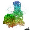 31225MC 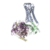 7eo4C 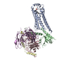 7wf7C M: map data used to model this data C: citing same article ( |
|---|---|
| Similar structure data |

-Links
 PDBj
PDBj


- Assembly
| Deposited unit | 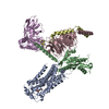
|
|---|---|
| 1 |
|
-Components
-Guanine nucleotide-binding protein ... , 3 types, 3 molecules BCD
| #2: Protein | Mass: 40445.059 Da / Num. of mol.: 1 Source method: isolated from a genetically manipulated source Source: (gene. exp.) Homo sapiens (human) / Gene: GNAI1 / Production host:  Insect BA phytoplasma (bacteria) / References: UniProt: P63096 Insect BA phytoplasma (bacteria) / References: UniProt: P63096 |
|---|---|
| #3: Protein | Mass: 37915.496 Da / Num. of mol.: 1 Source method: isolated from a genetically manipulated source Source: (gene. exp.) Homo sapiens (human) / Gene: GNB1 / Production host: Insect BA phytoplasma (bacteria) / References: UniProt: P62873 |
| #4: Protein | Mass: 7861.143 Da / Num. of mol.: 1 Source method: isolated from a genetically manipulated source Source: (gene. exp.) Homo sapiens (human) / Gene: GNG2 / Production host: Insect BA phytoplasma (bacteria) / References: UniProt: P59768 |
-Protein / Antibody / Non-polymers , 3 types, 3 molecules AE
| #1: Protein | Mass: 38240.746 Da / Num. of mol.: 1 Source method: isolated from a genetically manipulated source Source: (gene. exp.) Homo sapiens (human) / Gene: S1PR1, CHEDG1, EDG1 / Production host: Insect BA phytoplasma (bacteria) / References: UniProt: P21453 |
|---|---|
| #5: Antibody | Mass: 26293.299 Da / Num. of mol.: 1 Source method: isolated from a genetically manipulated source Source: (gene. exp.) Homo sapiens (human) / Production host: Insect BA phytoplasma (bacteria) |
| #6: Chemical | ChemComp-J89 / ( Mass: 387.451 Da / Num. of mol.: 1 / Source method: obtained synthetically / Formula: C19H34NO5P Mass: 387.451 Da / Num. of mol.: 1 / Source method: obtained synthetically / Formula: C19H34NO5P |
-Details
| Has ligand of interest | N |
|---|---|
| Has protein modification | Y |
-Experimental details
-Experiment
| Experiment | Method: ELECTRON MICROSCOPY |
|---|---|
| EM experiment | Aggregation state: 3D ARRAY / 3D reconstruction method: single particle reconstruction |
- Sample preparation
| Component | Name: S1PR1/Gi complex / Type: COMPLEX / Entity ID: #1-#5 / Source: RECOMBINANT |
|---|---|
| Source (natural) | Organism: Homo sapiens (human) |
| Source (recombinant) | Organism: Insect BA phytoplasma (bacteria) |
| Buffer solution | pH: 7.5 |
| Specimen | Embedding applied: NO / Shadowing applied: NO / Staining applied: NO / Vitrification applied: YES |
| Vitrification | Cryogen name: ETHANE |
- Electron microscopy imaging
| Experimental equipment |  Model: Titan Krios / Image courtesy: FEI Company |
|---|---|
| Microscopy | Model: FEI TITAN KRIOS |
| Electron gun | Electron source:  FIELD EMISSION GUN / Accelerating voltage: 300 kV / Illumination mode: OTHER FIELD EMISSION GUN / Accelerating voltage: 300 kV / Illumination mode: OTHER |
| Electron lens | Mode: BRIGHT FIELD |
| Image recording | Electron dose: 60 e/Å2 / Film or detector model: GATAN K2 SUMMIT (4k x 4k) |
| EM diffraction | Camera length: 800 mm |
| EM diffraction shell | Resolution: 3→5.5 Å / Fourier space coverage: 93.2 % / Multiplicity: 2.5 / Num. of structure factors: 244 / Phase residual: 13.5 ° |
| EM diffraction stats | Fourier space coverage: 90.3 % / High resolution: 2.83 Å / Num. of intensities measured: 1590 / Num. of structure factors: 325 / Phase error rejection criteria: 20 / Rmerge: 0.198 |
- Processing
| EM software |
| |||||||||
|---|---|---|---|---|---|---|---|---|---|---|
| CTF correction | Type: NONE | |||||||||
| 3D reconstruction | Resolution: 2.89 Å / Resolution method: FSC 0.143 CUT-OFF / Num. of particles: 600000 / Symmetry type: POINT | |||||||||
| Atomic model building | Protocol: OTHER / Space: REAL | |||||||||
| Atomic model building | PDB-ID: 6VMS Pdb chain-ID: A / Accession code: 6VMS / Source name: PDB / Type: experimental model |