Resolution: 2.703→38.093 Å / Cor.coef. Fo:Fc: 0.942 / Cor.coef. Fo:Fc free: 0.878 / SU B: 17.603 / SU ML: 0.355 / Cross valid method: FREE R-VALUE / ESU R Free: 0.448 Details: Hydrogens have been added in their riding positions
Rfactor
Num. reflection
% reflection
Rfree
0.293
2490
4.982 %
Rwork
0.198
47490
-
all
0.203
-
-
obs
-
49980
98.306 %
Solvent computation
Ion probe radii: 0.8 Å / Shrinkage radii: 0.8 Å / VDW probe radii: 1.2 Å / Solvent model: MASK BULK SOLVENT
Displacement parameters
Biso mean: 58.994 Å2
Baniso -1
Baniso -2
Baniso -3
1-
-0.028 Å2
0 Å2
-0.014 Å2
2-
-
0.019 Å2
0 Å2
3-
-
-
0.012 Å2
Refinement step
Cycle: LAST / Resolution: 2.703→38.093 Å
Protein
Nucleic acid
Ligand
Solvent
Total
Num. atoms
14381
0
0
147
14528
Refine LS restraints
Refine-ID
Type
Dev ideal
Dev ideal target
Number
X-RAY DIFFRACTION
r_bond_refined_d
0.008
0.013
14594
X-RAY DIFFRACTION
r_bond_other_d
0.001
0.017
14207
X-RAY DIFFRACTION
r_angle_refined_deg
1.579
1.621
19747
X-RAY DIFFRACTION
r_angle_other_deg
1.231
1.577
32638
X-RAY DIFFRACTION
r_dihedral_angle_1_deg
8.269
5
1952
X-RAY DIFFRACTION
r_dihedral_angle_2_deg
41.914
23.948
651
X-RAY DIFFRACTION
r_dihedral_angle_3_deg
19.059
15
2433
X-RAY DIFFRACTION
r_dihedral_angle_4_deg
16.272
15
65
X-RAY DIFFRACTION
r_chiral_restr
0.063
0.2
2031
X-RAY DIFFRACTION
r_gen_planes_refined
0.006
0.02
16939
X-RAY DIFFRACTION
r_gen_planes_other
0.001
0.02
3171
X-RAY DIFFRACTION
r_nbd_refined
0.213
0.2
3198
X-RAY DIFFRACTION
r_symmetry_nbd_other
0.186
0.2
14143
X-RAY DIFFRACTION
r_nbtor_refined
0.154
0.2
6992
X-RAY DIFFRACTION
r_symmetry_nbtor_other
0.079
0.2
7466
X-RAY DIFFRACTION
r_xyhbond_nbd_refined
0.175
0.2
403
X-RAY DIFFRACTION
r_symmetry_xyhbond_nbd_other
0.071
0.2
10
X-RAY DIFFRACTION
r_symmetry_nbd_refined
0.151
0.2
29
X-RAY DIFFRACTION
r_nbd_other
0.171
0.2
117
X-RAY DIFFRACTION
r_symmetry_xyhbond_nbd_refined
0.233
0.2
5
X-RAY DIFFRACTION
r_mcbond_it
4.999
6.105
7847
X-RAY DIFFRACTION
r_mcbond_other
4.99
6.105
7846
X-RAY DIFFRACTION
r_mcangle_it
7.563
9.136
9783
X-RAY DIFFRACTION
r_mcangle_other
7.562
9.136
9784
X-RAY DIFFRACTION
r_scbond_it
4.928
6.565
6747
X-RAY DIFFRACTION
r_scbond_other
4.928
6.566
6748
X-RAY DIFFRACTION
r_scangle_it
7.589
9.658
9963
X-RAY DIFFRACTION
r_scangle_other
7.588
9.659
9964
X-RAY DIFFRACTION
r_lrange_it
10.916
73.257
15565
X-RAY DIFFRACTION
r_lrange_other
10.915
73.263
15566
LS refinement shell
Resolution (Å)
Rfactor Rfree
Num. reflection Rfree
Rfactor Rwork
Num. reflection Rwork
Refine-ID
% reflection obs (%)
2.703-2.773
0.355
168
0.249
3402
X-RAY DIFFRACTION
96.1487
2.773-2.849
0.336
166
0.235
3481
X-RAY DIFFRACTION
100
2.849-2.932
0.324
177
0.227
3380
X-RAY DIFFRACTION
99.9719
2.932-3.022
0.344
163
0.214
3259
X-RAY DIFFRACTION
99.8832
3.022-3.121
0.314
162
0.223
3182
X-RAY DIFFRACTION
99.9402
3.121-3.23
0.341
161
0.214
3074
X-RAY DIFFRACTION
100
3.23-3.352
0.351
149
0.22
2934
X-RAY DIFFRACTION
99.8704
3.352-3.489
0.316
136
0.201
2892
X-RAY DIFFRACTION
99.7037
3.489-3.644
0.284
134
0.173
2739
X-RAY DIFFRACTION
99.6531
3.644-3.822
0.273
132
0.179
2607
X-RAY DIFFRACTION
99.5276
3.822-4.029
0.262
145
0.184
2469
X-RAY DIFFRACTION
99.3539
4.029-4.273
0.258
130
0.161
2321
X-RAY DIFFRACTION
98.7908
4.273-4.568
0.266
109
0.154
2170
X-RAY DIFFRACTION
97.1027
4.568-4.933
0.279
129
0.172
1987
X-RAY DIFFRACTION
96.8421
4.933-5.404
0.273
103
0.2
1841
X-RAY DIFFRACTION
97.4925
5.404-6.041
0.28
105
0.191
1688
X-RAY DIFFRACTION
98.5165
6.041-6.973
0.288
81
0.171
1518
X-RAY DIFFRACTION
98.948
6.973-8.537
0.247
72
0.19
1231
X-RAY DIFFRACTION
94.9017
8.537-12.055
0.283
37
0.246
830
X-RAY DIFFRACTION
80.6512
12.055-30.093
0.375
31
0.318
483
X-RAY DIFFRACTION
83.9869
+
About Yorodumi
-
News
-
Feb 9, 2022. New format data for meta-information of EMDB entries
New format data for meta-information of EMDB entries
Version 3 of the EMDB header file is now the official format.
The previous official version 1.9 will be removed from the archive.
In the structure databanks used in Yorodumi, some data are registered as the other names, "COVID-19 virus" and "2019-nCoV". Here are the details of the virus and the list of structure data.
Jan 31, 2019. EMDB accession codes are about to change! (news from PDBe EMDB page)
EMDB accession codes are about to change! (news from PDBe EMDB page)
The allocation of 4 digits for EMDB accession codes will soon come to an end. Whilst these codes will remain in use, new EMDB accession codes will include an additional digit and will expand incrementally as the available range of codes is exhausted. The current 4-digit format prefixed with “EMD-” (i.e. EMD-XXXX) will advance to a 5-digit format (i.e. EMD-XXXXX), and so on. It is currently estimated that the 4-digit codes will be depleted around Spring 2019, at which point the 5-digit format will come into force.
The EM Navigator/Yorodumi systems omit the EMD- prefix.
Related info.:Q: What is EMD? / ID/Accession-code notation in Yorodumi/EM Navigator
Yorodumi is a browser for structure data from EMDB, PDB, SASBDB, etc.
This page is also the successor to EM Navigator detail page, and also detail information page/front-end page for Omokage search.
The word "yorodu" (or yorozu) is an old Japanese word meaning "ten thousand". "mi" (miru) is to see.
Related info.:EMDB / PDB / SASBDB / Comparison of 3 databanks / Yorodumi Search / Aug 31, 2016. New EM Navigator & Yorodumi / Yorodumi Papers / Jmol/JSmol / Function and homology information / Changes in new EM Navigator and Yorodumi
 Movie
Movie Controller

 Yorodumi
 Open data
 Basic information
 Components
 Keywords
 Function and homology information
 X-RAY DIFFRACTION /
  Authors
 Citation
 Structure visualization
 Downloads & links
 Other downloads
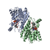


 PDBj
PDBj


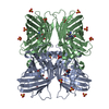
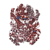
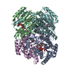
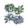
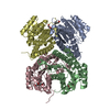
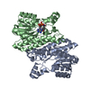
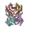
 Assembly
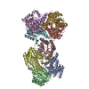

 Mass: 18.015 Da / Num. of mol.: 147 / Source method: isolated from a natural source / Formula: H2O
 Sample preparation
 / Beamline: 5C (4A) / Wavelength: 1 Å
 Processing