 Movie
Movie Controller


+ Open data

- Basic information
| Entry | Database: PDB / ID: 7emr | ||||||||||||
|---|---|---|---|---|---|---|---|---|---|---|---|---|---|
| Title | Crystal Structure of HasAp Capturing Cobalt Tetraphenylporphyrin | ||||||||||||
 Components Components | Heme acquisition protein HasAp | ||||||||||||
 Keywords Keywords | TRANSPORT PROTEIN / HEME ACQUISITION PROTEIN | ||||||||||||
| Function / homology | Haem-binding HasA / Haem-binding HasA superfamily / Heme-binding protein A (HasA) / metal ion binding / Co-5,10,15,20-Tetraphenylporphyrin / PHOSPHATE ION / Heme acquisition protein HasAp Function and homology information Function and homology information | ||||||||||||
| Biological species |  Pseudomonas aeruginosa str. PAO1 (bacteria) Pseudomonas aeruginosa str. PAO1 (bacteria) | ||||||||||||
| Method |  X-RAY DIFFRACTION / SYNCHROTRON / MOLECULAR REPLACEMENT / Resolution: 1.55 Å X-RAY DIFFRACTION / SYNCHROTRON / MOLECULAR REPLACEMENT / Resolution: 1.55 Å | ||||||||||||
 Authors Authors | Shisaka, Y. / Sakakibara, E. / Sugimoto, H. / Shoji, O. | ||||||||||||
| Funding support |  Japan, 3items Japan, 3items
| ||||||||||||
 Citation Citation | Journal: Chembiochem / Year: 2022 Title: Tetraphenylporphyrin Enters the Ring: First Example of a Complex between Highly Bulky Porphyrins and a Protein. Authors: Shisaka, Y. / Sakakibara, E. / Suzuki, K. / Stanfield, J.K. / Onoda, H. / Ueda, G. / Hatano, M. / Sugimoto, H. / Shoji, O. | ||||||||||||
| History |
|
- Structure visualization
| Structure viewer | Molecule: MolmilJmol/JSmol |
|---|
- Downloads & links
-Download
| PDBx/mmCIF format | 7emr.cif.gz | 240 KB | Display | PDBx/mmCIF format |
|---|---|---|---|---|
| PDB format | pdb7emr.ent.gz | 191.3 KB | Display | PDB format |
| PDBx/mmJSON format | 7emr.json.gz | Tree view | PDBx/mmJSON format | |
| Others |  Other downloads Other downloads |
-Validation report
| Summary document | 7emr_validation.pdf.gz | 1.5 MB | Display | wwPDB validaton report |
|---|---|---|---|---|
| Full document | 7emr_full_validation.pdf.gz | 1.5 MB | Display | |
| Data in XML | 7emr_validation.xml.gz | 25.7 KB | Display | |
| Data in CIF | 7emr_validation.cif.gz | 35.5 KB | Display | |
| Arichive directory | https://data.pdbj.org/pub/pdb/validation_reports/em/7emrftp://data.pdbj.org/pub/pdb/validation_reports/em/7emr | HTTPS FTP |
-Related structure data
| Related structure data |  7emoC 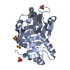 7empC 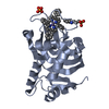 7emqC  7emsC  7emtC 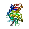 7emuC 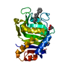 7emvC 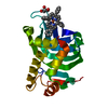 7emwC 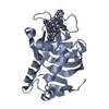 7vm1C 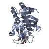 3ellS S: Starting model for refinement C: citing same article ( |
|---|---|
| Similar structure data |
-Links
 PDBj
PDBj- Assembly
| Deposited unit | 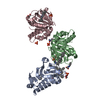
| ||||||||
|---|---|---|---|---|---|---|---|---|---|
| 1 | 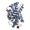
| ||||||||
| 2 | 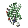
| ||||||||
| 3 | 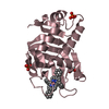
| ||||||||
| Unit cell |
|
-Components
-Protein , 1 types, 3 molecules ABC
| #1: Protein | Mass: 18901.535 Da / Num. of mol.: 3 Source method: isolated from a genetically manipulated source Source: (gene. exp.) Pseudomonas aeruginosa str. PAO1 (bacteria)Gene: hasAp, PA3407 / Plasmid: pQE30 / Production host: |
|---|
-Non-polymers , 6 types, 275 molecules 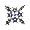


| #2: Chemical |  Mass: 671.653 Da / Num. of mol.: 3 / Source method: obtained synthetically / Formula: C44H28CoN4 / Feature type: SUBJECT OF INVESTIGATION Mass: 671.653 Da / Num. of mol.: 3 / Source method: obtained synthetically / Formula: C44H28CoN4 / Feature type: SUBJECT OF INVESTIGATION#3: Chemical | ChemComp-NHE / |  Mass: 207.290 Da / Num. of mol.: 1 / Source method: obtained synthetically / Formula: C8H17NO3S / Comment: pH buffer*YM Mass: 207.290 Da / Num. of mol.: 1 / Source method: obtained synthetically / Formula: C8H17NO3S / Comment: pH buffer*YM#4: Chemical |  Mass: 92.094 Da / Num. of mol.: 3 / Source method: obtained synthetically / Formula: C3H8O3 Mass: 92.094 Da / Num. of mol.: 3 / Source method: obtained synthetically / Formula: C3H8O3#5: Chemical | ChemComp-PO4 /  Mass: 94.971 Da / Num. of mol.: 5 / Source method: obtained synthetically / Formula: PO4 Mass: 94.971 Da / Num. of mol.: 5 / Source method: obtained synthetically / Formula: PO4#6: Chemical |  Mass: 221.317 Da / Num. of mol.: 3 / Source method: obtained synthetically / Formula: C9H19NO3S / Comment: pH buffer*YM Mass: 221.317 Da / Num. of mol.: 3 / Source method: obtained synthetically / Formula: C9H19NO3S / Comment: pH buffer*YM#7: Water | ChemComp-HOH / | Mass: 18.015 Da / Num. of mol.: 260 / Source method: isolated from a natural source / Formula: H2O |
|---|
-Details
| Has ligand of interest | Y |
|---|
-Experimental details
-Experiment
| Experiment | Method: X-RAY DIFFRACTION / Number of used crystals: 1 |
|---|
- Sample preparation
| Crystal | Density Matthews: 2.68 Å3/Da / Density % sol: 54.14 % / Mosaicity: 0.16 ° |
|---|---|
| Crystal grow | Temperature: 293 K / Method: vapor diffusion, sitting drop / pH: 10.5 Details: 100mM CAPS/NaOH (pH 10.5), 1.2M Sodium phosphate monobasic/0.8M pottasium phosphate dibasic, 0.2M lithium sulfate |
-Data collection
| Diffraction | Mean temperature: 100 K / Serial crystal experiment: N | ||||||||||||||||||||||||||||||
|---|---|---|---|---|---|---|---|---|---|---|---|---|---|---|---|---|---|---|---|---|---|---|---|---|---|---|---|---|---|---|---|
| Diffraction source | Source: SYNCHROTRON / Site: SPring-8 / Beamline: BL26B2 / Wavelength: 1 Å | ||||||||||||||||||||||||||||||
| Detector | Type: RAYONIX MX225-HS / Detector: CCD / Date: Dec 18, 2018 | ||||||||||||||||||||||||||||||
| Radiation | Monochromator: Si 111 CHANNEL / Protocol: SINGLE WAVELENGTH / Monochromatic (M) / Laue (L): M / Scattering type: x-ray | ||||||||||||||||||||||||||||||
| Radiation wavelength | Wavelength: 1 Å / Relative weight: 1 | ||||||||||||||||||||||||||||||
| Reflection | Resolution: 1.55→48.92 Å / Num. obs: 86763 / % possible obs: 100 % / Redundancy: 14.9 % / CC1/2: 1 / Rmerge(I) obs: 0.07 / Rpim(I) all: 0.019 / Rrim(I) all: 0.073 / Net I/σ(I): 22 / Num. measured all: 1295135 / Scaling rejects: 13 | ||||||||||||||||||||||||||||||
| Reflection shell | Diffraction-ID: 1
|
- Processing
| Software |
| |||||||||||||||||||||||||||||||||||||||||||||||||||||||||||||||||
|---|---|---|---|---|---|---|---|---|---|---|---|---|---|---|---|---|---|---|---|---|---|---|---|---|---|---|---|---|---|---|---|---|---|---|---|---|---|---|---|---|---|---|---|---|---|---|---|---|---|---|---|---|---|---|---|---|---|---|---|---|---|---|---|---|---|---|
| Refinement | Method to determine structure: MOLECULAR REPLACEMENT Starting model: 3ELL Resolution: 1.55→20 Å / Cor.coef. Fo:Fc: 0.978 / Cor.coef. Fo:Fc free: 0.969 / SU B: 2.695 / SU ML: 0.042 / Cross valid method: THROUGHOUT / σ(F): 0 / ESU R: 0.07 / ESU R Free: 0.061 / Stereochemistry target values: MAXIMUM LIKELIHOOD Details: HYDROGENS HAVE BEEN ADDED IN THE RIDING POSITIONS U VALUES : REFINED INDIVIDUALLY
| |||||||||||||||||||||||||||||||||||||||||||||||||||||||||||||||||
| Solvent computation | Ion probe radii: 0.8 Å / Shrinkage radii: 0.8 Å / VDW probe radii: 1.2 Å / Solvent model: MASK | |||||||||||||||||||||||||||||||||||||||||||||||||||||||||||||||||
| Displacement parameters | Biso max: 83.97 Å2 / Biso mean: 24.251 Å2 / Biso min: 15.19 Å2
| |||||||||||||||||||||||||||||||||||||||||||||||||||||||||||||||||
| Refinement step | Cycle: final / Resolution: 1.55→20 Å
| |||||||||||||||||||||||||||||||||||||||||||||||||||||||||||||||||
| Refine LS restraints |
| |||||||||||||||||||||||||||||||||||||||||||||||||||||||||||||||||
| LS refinement shell | Resolution: 1.55→1.59 Å / Rfactor Rfree error: 0 / Total num. of bins used: 20
|
