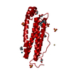
 Movie
Movie Controller


+ Open data

- Basic information
| Entry | Database: PDB / ID: 7eml | ||||||
|---|---|---|---|---|---|---|---|
| Title | Structure of IrCp* immobilized apo-D38H-rHLFr | ||||||
 Components Components | Ferritin light chain | ||||||
 Keywords Keywords | METAL BINDING PROTEIN / Iron storage protein / Ir binding | ||||||
| Function / homology |  Function and homology information Function and homology informationferritin complex / autolysosome / ferric iron binding / autophagosome / iron ion transport / ferrous iron binding / cytoplasmic vesicle / intracellular iron ion homeostasis / iron ion binding / cytoplasm Similarity search - Function | ||||||
| Biological species |  | ||||||
| Method |  X-RAY DIFFRACTION / SYNCHROTRON / MOLECULAR REPLACEMENT / Resolution: 1.25 Å X-RAY DIFFRACTION / SYNCHROTRON / MOLECULAR REPLACEMENT / Resolution: 1.25 Å | ||||||
 Authors Authors | Taher, M. / Maity, B. / Nakane, T. / Abe, S. / Ueno, T. / Mazumdar, S. | ||||||
| Funding support |  Japan, 1items Japan, 1items
| ||||||
 Citation Citation | Journal: Angew.Chem.Int.Ed.Engl. / Year: 2022 Title: Controlled Uptake of an Iridium Complex inside Engineered apo-Ferritin Nanocages: Study of Structure and Catalysis. Authors: Taher, M. / Maity, B. / Nakane, T. / Abe, S. / Ueno, T. / Mazumdar, S. | ||||||
| History |
|
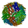
- Structure visualization
| Structure viewer | Molecule: MolmilJmol/JSmol |
|---|
- Downloads & links
-Download
| PDBx/mmCIF format | 7eml.cif.gz | 63.7 KB | Display | PDBx/mmCIF format |
|---|---|---|---|---|
| PDB format | pdb7eml.ent.gz | 44.2 KB | Display | PDB format |
| PDBx/mmJSON format | 7eml.json.gz | Tree view | PDBx/mmJSON format | |
| Others |  Other downloads Other downloads |
-Validation report
| Arichive directory | https://data.pdbj.org/pub/pdb/validation_reports/em/7emlftp://data.pdbj.org/pub/pdb/validation_reports/em/7eml | HTTPS FTP |
|---|
-Related structure data
| Related structure data |  7emmC  7w7jC 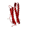 1datS S: Starting model for refinement C: citing same article ( |
|---|---|
| Similar structure data |


-Links
 PDBj
PDBj


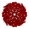
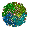
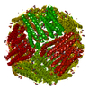
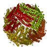
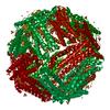
- Assembly
| Deposited unit | 
| |||||||||||||||
|---|---|---|---|---|---|---|---|---|---|---|---|---|---|---|---|---|
| 1 | x 24
| |||||||||||||||
| Unit cell |
| |||||||||||||||
| Components on special symmetry positions |
|
-Components
-Protein , 1 types, 1 molecules A
| #1: Protein | Mass: 19895.486 Da / Num. of mol.: 1 / Mutation: D38H Source method: isolated from a genetically manipulated source Source: (gene. exp.)  |
|---|
-Non-polymers , 7 types, 253 molecules 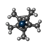


| #2: Chemical | ChemComp-RIR / [( Mass: 327.443 Da / Num. of mol.: 1 / Source method: obtained synthetically / Formula: C10H15Ir / Feature type: SUBJECT OF INVESTIGATION Mass: 327.443 Da / Num. of mol.: 1 / Source method: obtained synthetically / Formula: C10H15Ir / Feature type: SUBJECT OF INVESTIGATION | ||||||||||
|---|---|---|---|---|---|---|---|---|---|---|---|
| #3: Chemical |  Mass: 62.068 Da / Num. of mol.: 2 / Source method: obtained synthetically / Formula: C2H6O2 Mass: 62.068 Da / Num. of mol.: 2 / Source method: obtained synthetically / Formula: C2H6O2#4: Chemical |  Mass: 96.063 Da / Num. of mol.: 2 / Source method: obtained synthetically / Formula: SO4 Mass: 96.063 Da / Num. of mol.: 2 / Source method: obtained synthetically / Formula: SO4#5: Chemical | ChemComp-CD /  Mass: 112.411 Da / Num. of mol.: 5 / Source method: obtained synthetically / Formula: Cd Mass: 112.411 Da / Num. of mol.: 5 / Source method: obtained synthetically / Formula: Cd#6: Chemical | ChemComp-CL / |  Mass: 35.453 Da / Num. of mol.: 1 / Source method: obtained synthetically / Formula: Cl Mass: 35.453 Da / Num. of mol.: 1 / Source method: obtained synthetically / Formula: Cl#7: Chemical | ChemComp-IR /  Mass: 192.217 Da / Num. of mol.: 4 / Source method: obtained synthetically / Formula: Ir / Feature type: SUBJECT OF INVESTIGATION Mass: 192.217 Da / Num. of mol.: 4 / Source method: obtained synthetically / Formula: Ir / Feature type: SUBJECT OF INVESTIGATION#8: Water | ChemComp-HOH / | Mass: 18.015 Da / Num. of mol.: 238 / Source method: isolated from a natural source / Formula: H2O |
-Details
| Has ligand of interest | Y |
|---|
-Experimental details
-Experiment
| Experiment | Method: X-RAY DIFFRACTION / Number of used crystals: 1 |
|---|
- Sample preparation
| Crystal | Density Matthews: 3.19 Å3/Da / Density % sol: 61.5 % |
|---|---|
| Crystal grow | Temperature: 293 K / Method: vapor diffusion, hanging drop Details: Ammonium sulfate (0.5-1.0M), Cadmium sulfate(12-20mM) |
-Data collection
| Diffraction | Mean temperature: 100 K / Serial crystal experiment: N |
|---|---|
| Diffraction source | Source: SYNCHROTRON / Site: SPring-8 / Beamline: BL45XU / Wavelength: 1.05 Å |
| Detector | Type: DECTRIS PILATUS 6M / Detector: PIXEL / Date: Jun 20, 2019 |
| Radiation | Protocol: SINGLE WAVELENGTH / Monochromatic (M) / Laue (L): M / Scattering type: x-ray |
| Radiation wavelength | Wavelength: 1.05 Å / Relative weight: 1 |
| Reflection | Resolution: 1.25→45.526 Å / Num. obs: 71305 / % possible obs: 100 % / Redundancy: 72.5 % / CC1/2: 0.999 / Rmerge(I) obs: 0.2 / Net I/σ(I): 23.1 |
| Reflection shell | Resolution: 1.25→1.27 Å / Mean I/σ(I) obs: 2.5 / Num. unique obs: 71305 / CC1/2: 0.82 |
- Processing
| Software |
| |||||||||||||||||||||||||||||||||||||||||||||||||||||||||||||||||||||||||||||||||||||||||||||||||||||||||||||||||||||||||||||||||||||||||||||||||||||||||||||||||||||||||||||||||||||||||||||||||||||||||||||||||||||||||||||||||||||||
|---|---|---|---|---|---|---|---|---|---|---|---|---|---|---|---|---|---|---|---|---|---|---|---|---|---|---|---|---|---|---|---|---|---|---|---|---|---|---|---|---|---|---|---|---|---|---|---|---|---|---|---|---|---|---|---|---|---|---|---|---|---|---|---|---|---|---|---|---|---|---|---|---|---|---|---|---|---|---|---|---|---|---|---|---|---|---|---|---|---|---|---|---|---|---|---|---|---|---|---|---|---|---|---|---|---|---|---|---|---|---|---|---|---|---|---|---|---|---|---|---|---|---|---|---|---|---|---|---|---|---|---|---|---|---|---|---|---|---|---|---|---|---|---|---|---|---|---|---|---|---|---|---|---|---|---|---|---|---|---|---|---|---|---|---|---|---|---|---|---|---|---|---|---|---|---|---|---|---|---|---|---|---|---|---|---|---|---|---|---|---|---|---|---|---|---|---|---|---|---|---|---|---|---|---|---|---|---|---|---|---|---|---|---|---|---|---|---|---|---|---|---|---|---|---|---|---|---|---|---|---|---|---|
| Refinement | Method to determine structure: MOLECULAR REPLACEMENT Starting model: 1DAT Resolution: 1.25→41.739 Å / Cor.coef. Fo:Fc: 0.966 / Cor.coef. Fo:Fc free: 0.964 / WRfactor Rfree: 0.177 / WRfactor Rwork: 0.167 / SU B: 0.482 / SU ML: 0.021 / Average fsc free: 0.9589 / Average fsc work: 0.9613 / Cross valid method: FREE R-VALUE / ESU R: 0.037 / ESU R Free: 0.038 Details: Hydrogens have been added in their riding positions
| |||||||||||||||||||||||||||||||||||||||||||||||||||||||||||||||||||||||||||||||||||||||||||||||||||||||||||||||||||||||||||||||||||||||||||||||||||||||||||||||||||||||||||||||||||||||||||||||||||||||||||||||||||||||||||||||||||||||
| Solvent computation | Ion probe radii: 0.8 Å / Shrinkage radii: 0.8 Å / VDW probe radii: 1.2 Å / Solvent model: MASK BULK SOLVENT | |||||||||||||||||||||||||||||||||||||||||||||||||||||||||||||||||||||||||||||||||||||||||||||||||||||||||||||||||||||||||||||||||||||||||||||||||||||||||||||||||||||||||||||||||||||||||||||||||||||||||||||||||||||||||||||||||||||||
| Displacement parameters | Biso mean: 14.399 Å2
| |||||||||||||||||||||||||||||||||||||||||||||||||||||||||||||||||||||||||||||||||||||||||||||||||||||||||||||||||||||||||||||||||||||||||||||||||||||||||||||||||||||||||||||||||||||||||||||||||||||||||||||||||||||||||||||||||||||||
| Refinement step | Cycle: LAST / Resolution: 1.25→41.739 Å
| |||||||||||||||||||||||||||||||||||||||||||||||||||||||||||||||||||||||||||||||||||||||||||||||||||||||||||||||||||||||||||||||||||||||||||||||||||||||||||||||||||||||||||||||||||||||||||||||||||||||||||||||||||||||||||||||||||||||
| Refine LS restraints |
| |||||||||||||||||||||||||||||||||||||||||||||||||||||||||||||||||||||||||||||||||||||||||||||||||||||||||||||||||||||||||||||||||||||||||||||||||||||||||||||||||||||||||||||||||||||||||||||||||||||||||||||||||||||||||||||||||||||||
| LS refinement shell | Refine-ID: X-RAY DIFFRACTION / Total num. of bins used: 20
|
