 Movie
Movie Controller


+ Open data

- Basic information
| Entry | Database: PDB / ID: 7emk | ||||||
|---|---|---|---|---|---|---|---|
| Title | Dendrorhynchus zhejiangensis ferritin | ||||||
 Components Components | Dendrorhynchus zhejiangensis ferritin | ||||||
 Keywords Keywords | STRUCTURAL PROTEIN / Dendrorhynchus zhejiangensis / Ferritin | ||||||
| Function / homology | Ferritin, core subunit, four-helix bundle / Ferritin / Up-down Bundle / Mainly Alpha / : / DI(HYDROXYETHYL)ETHER Function and homology information Function and homology information | ||||||
| Biological species |  Dendrorhynchus sinensis (invertebrata) Dendrorhynchus sinensis (invertebrata) | ||||||
| Method |  X-RAY DIFFRACTION / SYNCHROTRON / SAD / Resolution: 2.3 Å X-RAY DIFFRACTION / SYNCHROTRON / SAD / Resolution: 2.3 Å | ||||||
 Authors Authors | Huan, H.S. / Ming, T.H. / Su, X.R. | ||||||
| Funding support |  China, 1items China, 1items
| ||||||
 Citation Citation | Journal: Biochemical and Biophysical Research Communications Year: 2020 Title: Structure determination of ferritin from Dendrorhynchus zhejiangensis Authors: Huan, H.S. / Jiang, Q.Q. / Wu, Y. / Qiu, X.T. / Lu, C.Y. / Su, C. / Zhou, J. / Li, Y. / Ming, T.H. / Su, X.R. | ||||||
| History |
|
- Structure visualization
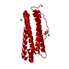
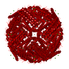
| Structure viewer | Molecule: MolmilJmol/JSmol |
|---|
- Downloads & links
-Download
| PDBx/mmCIF format | 7emk.cif.gz | 53.7 KB | Display | PDBx/mmCIF format |
|---|---|---|---|---|
| PDB format | pdb7emk.ent.gz | 37.6 KB | Display | PDB format |
| PDBx/mmJSON format | 7emk.json.gz | Tree view | PDBx/mmJSON format | |
| Others |  Other downloads Other downloads |
-Validation report
| Arichive directory | https://data.pdbj.org/pub/pdb/validation_reports/em/7emkftp://data.pdbj.org/pub/pdb/validation_reports/em/7emk | HTTPS FTP |
|---|
-Related structure data
| Similar structure data |
|---|


-Links
 PDBj
PDBj
- Assembly
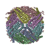
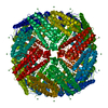
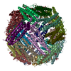
| Deposited unit | 
| |||||||||||||||
|---|---|---|---|---|---|---|---|---|---|---|---|---|---|---|---|---|
| 1 | x 24
| |||||||||||||||
| Unit cell |
| |||||||||||||||
| Components on special symmetry positions |
|
-Components
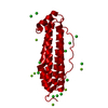
-Protein , 1 types, 1 molecules A
| #1: Protein | Mass: 19521.992 Da / Num. of mol.: 1 Source method: isolated from a genetically manipulated source Source: (gene. exp.) Dendrorhynchus sinensis (invertebrata) / Production host:  |
|---|
-Non-polymers , 5 types, 118 molecules 


| #2: Chemical | ChemComp-FE /  Mass: 55.845 Da / Num. of mol.: 8 / Source method: obtained synthetically / Formula: Fe / Feature type: SUBJECT OF INVESTIGATION Mass: 55.845 Da / Num. of mol.: 8 / Source method: obtained synthetically / Formula: Fe / Feature type: SUBJECT OF INVESTIGATION#3: Chemical | ChemComp-NA /  Mass: 22.990 Da / Num. of mol.: 17 / Source method: obtained synthetically / Formula: Na Mass: 22.990 Da / Num. of mol.: 17 / Source method: obtained synthetically / Formula: Na#4: Chemical | ChemComp-CL /  Mass: 35.453 Da / Num. of mol.: 21 / Source method: obtained synthetically / Formula: Cl Mass: 35.453 Da / Num. of mol.: 21 / Source method: obtained synthetically / Formula: Cl#5: Chemical | ChemComp-PEG / |  Mass: 106.120 Da / Num. of mol.: 1 / Source method: obtained synthetically / Formula: C4H10O3 Mass: 106.120 Da / Num. of mol.: 1 / Source method: obtained synthetically / Formula: C4H10O3#6: Water | ChemComp-HOH / | Mass: 18.015 Da / Num. of mol.: 71 / Source method: isolated from a natural source / Formula: H2O |
|---|
-Details
| Has ligand of interest | Y |
|---|
-Experimental details
-Experiment
| Experiment | Method: X-RAY DIFFRACTION / Number of used crystals: 1 |
|---|
- Sample preparation
| Crystal | Density Matthews: 3.73 Å3/Da / Density % sol: 67.04 % |
|---|---|
| Crystal grow | Temperature: 291.15 K / Method: vapor diffusion / pH: 7.5 Details: 0.2 M calcium chloride dihydrate, 0.1 M HEPES sodium, pH 7.5, and 28% (v/v) polyethylene glycol 400 |
-Data collection
| Diffraction | Mean temperature: 100 K / Serial crystal experiment: N | |||||||||||||||||||||||||||||||||||||||||||||||||||||||||||||||||||||||||||||||||||||||||||||||||||||||||||||||||||||||||||||||||||||||||||||||||||||||||||||||||||||||||||||||||||||||||||||
|---|---|---|---|---|---|---|---|---|---|---|---|---|---|---|---|---|---|---|---|---|---|---|---|---|---|---|---|---|---|---|---|---|---|---|---|---|---|---|---|---|---|---|---|---|---|---|---|---|---|---|---|---|---|---|---|---|---|---|---|---|---|---|---|---|---|---|---|---|---|---|---|---|---|---|---|---|---|---|---|---|---|---|---|---|---|---|---|---|---|---|---|---|---|---|---|---|---|---|---|---|---|---|---|---|---|---|---|---|---|---|---|---|---|---|---|---|---|---|---|---|---|---|---|---|---|---|---|---|---|---|---|---|---|---|---|---|---|---|---|---|---|---|---|---|---|---|---|---|---|---|---|---|---|---|---|---|---|---|---|---|---|---|---|---|---|---|---|---|---|---|---|---|---|---|---|---|---|---|---|---|---|---|---|---|---|---|---|---|---|---|
| Diffraction source | Source: SYNCHROTRON / Site: NFPSS / Beamline: BL18U / Wavelength: 0.96003 Å | |||||||||||||||||||||||||||||||||||||||||||||||||||||||||||||||||||||||||||||||||||||||||||||||||||||||||||||||||||||||||||||||||||||||||||||||||||||||||||||||||||||||||||||||||||||||||||||
| Detector | Type: DECTRIS PILATUS3 S 6M / Detector: PIXEL / Date: Dec 23, 2018 | |||||||||||||||||||||||||||||||||||||||||||||||||||||||||||||||||||||||||||||||||||||||||||||||||||||||||||||||||||||||||||||||||||||||||||||||||||||||||||||||||||||||||||||||||||||||||||||
| Radiation | Protocol: SINGLE WAVELENGTH / Monochromatic (M) / Laue (L): M / Scattering type: x-ray | |||||||||||||||||||||||||||||||||||||||||||||||||||||||||||||||||||||||||||||||||||||||||||||||||||||||||||||||||||||||||||||||||||||||||||||||||||||||||||||||||||||||||||||||||||||||||||||
| Radiation wavelength | Wavelength: 0.96003 Å / Relative weight: 1 | |||||||||||||||||||||||||||||||||||||||||||||||||||||||||||||||||||||||||||||||||||||||||||||||||||||||||||||||||||||||||||||||||||||||||||||||||||||||||||||||||||||||||||||||||||||||||||||
| Reflection | Resolution: 2.3→50 Å / Num. obs: 13520 / % possible obs: 100 % / Redundancy: 38 % / Rmerge(I) obs: 0.153 / Rpim(I) all: 0.025 / Rrim(I) all: 0.155 / Χ2: 1.124 / Net I/σ(I): 7.2 / Num. measured all: 513539 | |||||||||||||||||||||||||||||||||||||||||||||||||||||||||||||||||||||||||||||||||||||||||||||||||||||||||||||||||||||||||||||||||||||||||||||||||||||||||||||||||||||||||||||||||||||||||||||
| Reflection shell | Diffraction-ID: 1
|
- Processing
| Software |
| ||||||||||||||||||||||||||||||||||||||||||||||||||||||||||||
|---|---|---|---|---|---|---|---|---|---|---|---|---|---|---|---|---|---|---|---|---|---|---|---|---|---|---|---|---|---|---|---|---|---|---|---|---|---|---|---|---|---|---|---|---|---|---|---|---|---|---|---|---|---|---|---|---|---|---|---|---|---|
| Refinement | Method to determine structure: SAD / Resolution: 2.3→48 Å / Cor.coef. Fo:Fc: 0.965 / Cor.coef. Fo:Fc free: 0.947 / SU B: 3.863 / SU ML: 0.094 / Cross valid method: THROUGHOUT / σ(F): 0 / ESU R: 0.165 / ESU R Free: 0.15 / Stereochemistry target values: MAXIMUM LIKELIHOOD Details: HYDROGENS HAVE BEEN ADDED IN THE RIDING POSITIONS U VALUES : REFINED INDIVIDUALLY
| ||||||||||||||||||||||||||||||||||||||||||||||||||||||||||||
| Solvent computation | Ion probe radii: 0.8 Å / Shrinkage radii: 0.8 Å / VDW probe radii: 1.2 Å / Solvent model: MASK | ||||||||||||||||||||||||||||||||||||||||||||||||||||||||||||
| Displacement parameters | Biso max: 112.3 Å2 / Biso mean: 30.451 Å2 / Biso min: 18.47 Å2
| ||||||||||||||||||||||||||||||||||||||||||||||||||||||||||||
| Refinement step | Cycle: final / Resolution: 2.3→48 Å
| ||||||||||||||||||||||||||||||||||||||||||||||||||||||||||||
| Refine LS restraints |
| ||||||||||||||||||||||||||||||||||||||||||||||||||||||||||||
| LS refinement shell | Resolution: 2.305→2.364 Å / Rfactor Rfree error: 0 / Total num. of bins used: 20
|
