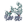
 Movie
Movie Controller


+ Open data

- Basic information
| Entry | Database: PDB / ID: 7el1 | |||||||||
|---|---|---|---|---|---|---|---|---|---|---|
| Title | Structure of a protein from bacteria | |||||||||
 Components Components |
| |||||||||
 Keywords Keywords | UNKNOWN FUNCTION | |||||||||
| Function / homology |  Function and homology information Function and homology informationmaintenance of CRISPR repeat elements / endonuclease activity / defense response to virus / Hydrolases; Acting on ester bonds / hydrolase activity / DNA binding / RNA binding / metal ion binding Similarity search - Function | |||||||||
| Biological species |   Staphylococcus aureus (bacteria) Staphylococcus aureus (bacteria) | |||||||||
| Method |  X-RAY DIFFRACTION / SYNCHROTRON / MOLECULAR REPLACEMENT / Resolution: 2.22 Å X-RAY DIFFRACTION / SYNCHROTRON / MOLECULAR REPLACEMENT / Resolution: 2.22 Å | |||||||||
 Authors Authors | Liu, H. / Zhu, Y. / Huang, Z. | |||||||||
| Funding support |  China, 2items China, 2items
| |||||||||
 Citation Citation | Journal: Nucleic Acids Res. / Year: 2021 Title: Structural basis of Staphylococcus aureus Cas9 inhibition by AcrIIA14. Authors: Liu, H. / Zhu, Y. / Lu, Z. / Huang, Z. | |||||||||
| History |
|
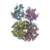
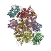
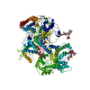
- Structure visualization
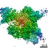
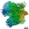
| Structure viewer | Molecule: MolmilJmol/JSmol |
|---|
- Downloads & links
-Download
| PDBx/mmCIF format | 7el1.cif.gz | 300.3 KB | Display | PDBx/mmCIF format |
|---|---|---|---|---|
| PDB format | pdb7el1.ent.gz | 233.6 KB | Display | PDB format |
| PDBx/mmJSON format | 7el1.json.gz | Tree view | PDBx/mmJSON format | |
| Others |  Other downloads Other downloads |
-Validation report
| Arichive directory | https://data.pdbj.org/pub/pdb/validation_reports/el/7el1ftp://data.pdbj.org/pub/pdb/validation_reports/el/7el1 | HTTPS FTP |
|---|
-Related structure data
| Related structure data |  5czzS S: Starting model for refinement |
|---|---|
| Similar structure data |

-Links
 PDBj
PDBj


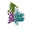
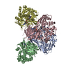
- Assembly
| Deposited unit | 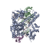
| ||||||||
|---|---|---|---|---|---|---|---|---|---|
| 1 |
| ||||||||
| Unit cell |
|
-Components
-Protein , 2 types, 2 molecules AE
| #1: Protein | Mass: 124083.648 Da / Num. of mol.: 1 / Mutation: N580A, C946A Source method: isolated from a genetically manipulated source Source: (gene. exp.) Staphylococcus aureus (bacteria) / Gene: cas9 / Production host: References: UniProt: J7RUA5, Hydrolases; Acting on ester bonds |
|---|---|
| #5: Protein | Mass: 11937.598 Da / Num. of mol.: 1 Source method: isolated from a genetically manipulated source Source: (gene. exp.) Staphylococcus aureus (bacteria) / Production host: |
-DNA chain , 2 types, 2 molecules CD
| #3: DNA chain | Mass: 8469.482 Da / Num. of mol.: 1 / Source method: obtained synthetically / Source: (synth.) Staphylococcus aureus (bacteria) |
|---|---|
| #4: DNA chain | Mass: 2465.653 Da / Num. of mol.: 1 / Source method: obtained synthetically / Source: (synth.) Staphylococcus aureus (bacteria) |
-RNA chain / Non-polymers , 2 types, 127 molecules B
| #2: RNA chain | Mass: 23525.975 Da / Num. of mol.: 1 / Source method: obtained synthetically / Source: (synth.) Staphylococcus aureus (bacteria) |
|---|---|
| #6: Water | ChemComp-HOH / Mass: 18.015 Da / Num. of mol.: 126 / Source method: isolated from a natural source / Formula: H2O |
-Experimental details
-Experiment
| Experiment | Method: X-RAY DIFFRACTION / Number of used crystals: 1 |
|---|
- Sample preparation
| Crystal | Density Matthews: 3.49 Å3/Da / Density % sol: 64.79 % |
|---|---|
| Crystal grow | Temperature: 293 K / Method: vapor diffusion, hanging drop Details: 1.0M sodium citrate dihydrate, 0.3M imidazol-Hcl (pH 10.0), 0.02M sodium malonate (pH 7.0) |
-Data collection
| Diffraction | Mean temperature: 100 K / Serial crystal experiment: N |
|---|---|
| Diffraction source | Source: SYNCHROTRON / Site: APS  / Beamline: 17-BM / Wavelength: 0.9791 Å / Beamline: 17-BM / Wavelength: 0.9791 Å |
| Detector | Type: DECTRIS EIGER X 16M / Detector: PIXEL / Date: Jan 18, 2021 |
| Radiation | Protocol: SINGLE WAVELENGTH / Monochromatic (M) / Laue (L): M / Scattering type: x-ray |
| Radiation wavelength | Wavelength: 0.9791 Å / Relative weight: 1 |
| Reflection | Resolution: 2.22→68.47 Å / Num. obs: 116344 / % possible obs: 99.9 % / Redundancy: 6.4 % / Rmerge(I) obs: 0.052 / Net I/σ(I): 17 |
| Reflection shell | Resolution: 2.22→2.34 Å / Rmerge(I) obs: 0.714 / Num. unique obs: 16935 |
- Processing
| Software |
| ||||||||||||||||
|---|---|---|---|---|---|---|---|---|---|---|---|---|---|---|---|---|---|
| Refinement | Method to determine structure: MOLECULAR REPLACEMENT Starting model: 5czz Resolution: 2.22→50 Å / Cross valid method: FREE R-VALUE
| ||||||||||||||||
| Refinement step | Cycle: LAST / Resolution: 2.22→50 Å
| ||||||||||||||||
| LS refinement shell | Resolution: 2.22→2.34 Å /
|
