 Movie
Movie Controller

[English] 日本語
 Yorodumi
- PDB-7eiq: Crystal structure of chondroitin ABC lyase I in complex with chon... -
+ Open data

- Basic information
| Entry | Database: PDB / ID: 7eiq | ||||||
|---|---|---|---|---|---|---|---|
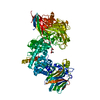
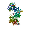
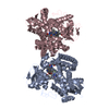
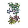
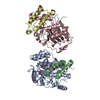
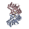
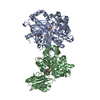
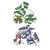
| Title | Crystal structure of chondroitin ABC lyase I in complex with chondroitin disaccharide 4S | ||||||
 Components Components | Chondroitin sulfate ABC endolyase | ||||||
 Keywords Keywords | LYASE / Polysaccharide lyase family 8 / carbohydrate binding | ||||||
| Function / homology |  Function and homology information Function and homology informationchondroitin-sulfate-ABC endolyase / chondroitin-sulfate-ABC endolyase activity / glycosaminoglycan catabolic process / carbohydrate binding / carbohydrate metabolic process / periplasmic space / extracellular region / metal ion binding Similarity search - Function | ||||||
| Biological species |  Proteus vulgaris (bacteria) Proteus vulgaris (bacteria) | ||||||
| Method |  X-RAY DIFFRACTION / SYNCHROTRON / MOLECULAR REPLACEMENT / Resolution: 1.8 Å X-RAY DIFFRACTION / SYNCHROTRON / MOLECULAR REPLACEMENT / Resolution: 1.8 Å | ||||||
 Authors Authors | Takashima, M. / Miyanaga, A. / Eguchi, T. | ||||||
 Citation Citation | Journal: Glycobiology / Year: 2021 Title: Substrate specificity of Chondroitinase ABC I based on analyses of biochemical reactions and crystal structures in complex with disaccharides. Authors: Takashima, M. / Watanabe, I. / Miyanaga, A. / Eguchi, T. | ||||||
| History |
|
- Structure visualization
| Structure viewer | Molecule: MolmilJmol/JSmol |
|---|
- Downloads & links
-Download
| PDBx/mmCIF format | 7eiq.cif.gz | 409.3 KB | Display | PDBx/mmCIF format |
|---|---|---|---|---|
| PDB format | pdb7eiq.ent.gz | 331.3 KB | Display | PDB format |
| PDBx/mmJSON format | 7eiq.json.gz | Tree view | PDBx/mmJSON format | |
| Others |  Other downloads Other downloads |
-Validation report
| Arichive directory | https://data.pdbj.org/pub/pdb/validation_reports/ei/7eiqftp://data.pdbj.org/pub/pdb/validation_reports/ei/7eiq | HTTPS FTP |
|---|
-Related structure data
| Related structure data |  7eipC 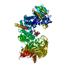 7eirC  7eisC  1hn0S S: Starting model for refinement C: citing same article ( |
|---|---|
| Similar structure data |
-Links
 PDBj
PDBj


- Assembly
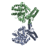
| Deposited unit | 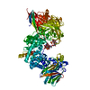
| ||||||||
|---|---|---|---|---|---|---|---|---|---|
| 1 |
| ||||||||
| Unit cell |
|
-Components
| #1: Protein | Mass: 115216.391 Da / Num. of mol.: 1 / Source method: isolated from a natural source / Source: (natural) Proteus vulgaris (bacteria)References: UniProt: P59807, chondroitin-sulfate-ABC endolyase | ||||||
|---|---|---|---|---|---|---|---|
| #2: Polysaccharide | Source method: isolated from a genetically manipulated source #3: Chemical | ChemComp-MG / |   Mass: 24.305 Da / Num. of mol.: 1 / Source method: obtained synthetically / Formula: Mg Mass: 24.305 Da / Num. of mol.: 1 / Source method: obtained synthetically / Formula: Mg#4: Water | ChemComp-HOH / |  Mass: 18.015 Da / Num. of mol.: 823 / Source method: isolated from a natural source / Formula: H2O Mass: 18.015 Da / Num. of mol.: 823 / Source method: isolated from a natural source / Formula: H2OHas ligand of interest | Y | |
-Experimental details
-Experiment
| Experiment | Method: X-RAY DIFFRACTION / Number of used crystals: 1 |
|---|
- Sample preparation
| Crystal | Density Matthews: 2.32 Å3/Da / Density % sol: 46.91 % |
|---|---|
| Crystal grow | Temperature: 293 K / Method: vapor diffusion, sitting drop / pH: 7.5 Details: magnesium acetate, ammonium acetate, polyethylene glycol 3350, HEPES-Na |
-Data collection
| Diffraction | Mean temperature: 100 K / Serial crystal experiment: N |
|---|---|
| Diffraction source | Source: SYNCHROTRON / Site: Photon Factory  / Beamline: AR-NW12A / Wavelength: 1 Å / Beamline: AR-NW12A / Wavelength: 1 Å |
| Detector | Type: DECTRIS PILATUS3 S 2M / Detector: PIXEL / Date: Nov 7, 2020 |
| Radiation | Monochromator: Numerical link type Si(111) double crystal monochromator Protocol: SINGLE WAVELENGTH / Monochromatic (M) / Laue (L): M / Scattering type: x-ray |
| Radiation wavelength | Wavelength: 1 Å / Relative weight: 1 |
| Reflection | Resolution: 1.8→50 Å / Num. obs: 100438 / % possible obs: 100 % / Redundancy: 6.5 % / CC1/2: 0.997 / Rmerge(I) obs: 0.101 / Net I/σ(I): 11.3 |
| Reflection shell | Resolution: 1.8→1.83 Å / Redundancy: 6.1 % / Rmerge(I) obs: 0.814 / Mean I/σ(I) obs: 2 / Num. unique obs: 4953 / CC1/2: 0.711 / % possible all: 100 |
- Processing
| Software |
| ||||||||||||||||||||||||||||||||||||||||||||||||||||||||||||
|---|---|---|---|---|---|---|---|---|---|---|---|---|---|---|---|---|---|---|---|---|---|---|---|---|---|---|---|---|---|---|---|---|---|---|---|---|---|---|---|---|---|---|---|---|---|---|---|---|---|---|---|---|---|---|---|---|---|---|---|---|---|
| Refinement | Method to determine structure: MOLECULAR REPLACEMENT Starting model: 1HN0 Resolution: 1.8→49.05 Å / Cor.coef. Fo:Fc: 0.964 / Cor.coef. Fo:Fc free: 0.942 / SU B: 5.812 / SU ML: 0.091 / Cross valid method: THROUGHOUT / σ(F): 0 / ESU R: 0.118 / ESU R Free: 0.118 / Stereochemistry target values: MAXIMUM LIKELIHOOD Details: U VALUES : WITH TLS ADDED HYDROGENS HAVE BEEN ADDED IN THE RIDING POSITIONS
| ||||||||||||||||||||||||||||||||||||||||||||||||||||||||||||
| Solvent computation | Ion probe radii: 0.8 Å / Shrinkage radii: 0.8 Å / VDW probe radii: 1.2 Å / Solvent model: MASK | ||||||||||||||||||||||||||||||||||||||||||||||||||||||||||||
| Displacement parameters | Biso max: 82.59 Å2 / Biso mean: 27.233 Å2 / Biso min: 11.07 Å2
| ||||||||||||||||||||||||||||||||||||||||||||||||||||||||||||
| Refinement step | Cycle: final / Resolution: 1.8→49.05 Å
| ||||||||||||||||||||||||||||||||||||||||||||||||||||||||||||
| Refine LS restraints |
| ||||||||||||||||||||||||||||||||||||||||||||||||||||||||||||
| LS refinement shell | Resolution: 1.8→1.847 Å / Rfactor Rfree error: 0 / Total num. of bins used: 20
| ||||||||||||||||||||||||||||||||||||||||||||||||||||||||||||
| Refinement TLS params. | Method: refined / Origin x: -8.587 Å / Origin y: 25.144 Å / Origin z: -31.967 Å
|
