- PDB-1hn0: CRYSTAL STRUCTURE OF CHONDROITIN ABC LYASE I FROM PROTEUS VULGARI... -
+
Open data
ID or keywords:
Loading...
-
Basic information
Entry
Database: PDB / ID: 1hn0
Title
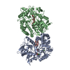
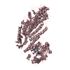
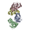
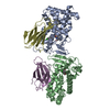
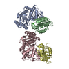
CRYSTAL STRUCTURE OF CHONDROITIN ABC LYASE I FROM PROTEUS VULGARIS AT 1.9 ANGSTROMS RESOLUTION
Components
CHONDROITIN ABC LYASE I
Keywords
LYASE / chondroitinase ABC I / chonroitin digestion
Function / homology
Function and homology information
chondroitin-sulfate-ABC endolyase / chondroitin-sulfate-ABC endolyase activity / glycosaminoglycan catabolic process / carbohydrate binding / carbohydrate metabolic process / periplasmic space / extracellular region / metal ion binding Similarity search - Function
Method to determine structure: MAD, MIR / Resolution: 1.9→19.88 Å / SU B: 3.024 / SU ML: 0.09 / Cross valid method: THROUGHOUT / ESU R: 0.149 / ESU R Free: 0.144 / Stereochemistry target values: MAXIMUM LIKELIHOOD
Rfactor
Num. reflection
% reflection
Selection details
Rfree
0.21224
1548
2 %
RANDOM
Rwork
0.15642
-
-
-
obs
0.15753
75799
100 %
-
Solvent computation
Ion probe radii: 0.8 Å / Shrinkage radii: 0.8 Å / VDW probe radii: 1.4 Å / Solvent model: BABINET MODEL WITH MASK
Displacement parameters
Biso mean: 25.781 Å2
Baniso -1
Baniso -2
Baniso -3
1-
0.7 Å2
0 Å2
0 Å2
2-
-
0.19 Å2
0 Å2
3-
-
-
-0.89 Å2
Refinement step
Cycle: LAST / Resolution: 1.9→19.88 Å
Protein
Nucleic acid
Ligand
Solvent
Total
Num. atoms
7759
0
1
1666
9426
Refine LS restraints
Refine-ID
Type
Dev ideal
Dev ideal target
Number
X-RAY DIFFRACTION
r_bond_refined_d
0.016
0.021
7953
X-RAY DIFFRACTION
r_bond_other_d
X-RAY DIFFRACTION
r_angle_refined_deg
1.443
1.934
10789
X-RAY DIFFRACTION
r_angle_other_deg
X-RAY DIFFRACTION
r_dihedral_angle_1_deg
6.657
5
969
X-RAY DIFFRACTION
r_dihedral_angle_3_deg
15.891
15
1356
X-RAY DIFFRACTION
r_chiral_restr
0.107
0.2
1165
X-RAY DIFFRACTION
r_gen_planes_refined
0.007
0.02
6082
X-RAY DIFFRACTION
r_gen_planes_other
X-RAY DIFFRACTION
r_nbd_refined
0.212
0.2
4007
X-RAY DIFFRACTION
r_nbd_other
X-RAY DIFFRACTION
r_nbtor_other
X-RAY DIFFRACTION
r_xyhbond_nbd_refined
0.178
0.2
1360
X-RAY DIFFRACTION
r_xyhbond_nbd_other
X-RAY DIFFRACTION
r_symmetry_vdw_refined
0.202
0.2
69
X-RAY DIFFRACTION
r_symmetry_vdw_other
X-RAY DIFFRACTION
r_symmetry_hbond_refined
0.197
0.2
113
X-RAY DIFFRACTION
r_symmetry_hbond_other
X-RAY DIFFRACTION
r_mcbond_it
0.847
1.5
4841
X-RAY DIFFRACTION
r_mcangle_it
1.507
2
7821
X-RAY DIFFRACTION
r_scbond_it
2.403
3
3112
X-RAY DIFFRACTION
r_scangle_it
3.728
4.5
2968
X-RAY DIFFRACTION
r_rigid_bond_restr
X-RAY DIFFRACTION
r_sphericity_free
X-RAY DIFFRACTION
r_sphericity_bonded
LS refinement shell
Resolution: 1.905→1.954 Å / Total num. of bins used: 20 /
In the structure databanks used in Yorodumi, some data are registered as the other names, "COVID-19 virus" and "2019-nCoV". Here are the details of the virus and the list of structure data.
Jan 31, 2019. EMDB accession codes are about to change! (news from PDBe EMDB page)
EMDB accession codes are about to change! (news from PDBe EMDB page)
The allocation of 4 digits for EMDB accession codes will soon come to an end. Whilst these codes will remain in use, new EMDB accession codes will include an additional digit and will expand incrementally as the available range of codes is exhausted. The current 4-digit format prefixed with “EMD-” (i.e. EMD-XXXX) will advance to a 5-digit format (i.e. EMD-XXXXX), and so on. It is currently estimated that the 4-digit codes will be depleted around Spring 2019, at which point the 5-digit format will come into force.
The EM Navigator/Yorodumi systems omit the EMD- prefix.
Related info.:Q: What is EMD? / ID/Accession-code notation in Yorodumi/EM Navigator
Yorodumi is a browser for structure data from EMDB, PDB, SASBDB, etc.
This page is also the successor to EM Navigator detail page, and also detail information page/front-end page for Omokage search.
The word "yorodu" (or yorozu) is an old Japanese word meaning "ten thousand". "mi" (miru) is to see.
Related info.:EMDB / PDB / SASBDB / Comparison of 3 databanks / Yorodumi Search / Aug 31, 2016. New EM Navigator & Yorodumi / Yorodumi Papers / Jmol/JSmol / Function and homology information / Changes in new EM Navigator and Yorodumi
 Movie
Movie Controller

 Yorodumi
 Open data
 Basic information
 Components
 Keywords
 Function and homology information
 Proteus vulgaris (bacteria)
 X-RAY DIFFRACTION /
  Authors
 Citation
 Structure visualization
 Downloads & links
 Other downloads
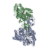


 PDBj
PDBj


 Assembly
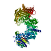

 Mass: 22.990 Da / Num. of mol.: 1 / Source method: obtained synthetically / Formula: Na
 Mass: 18.015 Da / Num. of mol.: 1666 / Source method: isolated from a natural source / Formula: H2O
 Sample preparation
 / Beamline: X8C / Wavelength: 1.006 Å
 Processing