 Movie
Movie Controller


+ Open data

- Basic information
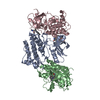
| Entry | Database: PDB / ID: 1f3m | ||||||
|---|---|---|---|---|---|---|---|
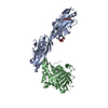
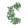
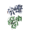
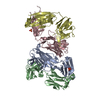
| Title | CRYSTAL STRUCTURE OF HUMAN SERINE/THREONINE KINASE PAK1 | ||||||
 Components Components | (SERINE/THREONINE-PROTEIN KINASE PAK-ALPHA) x 2 | ||||||
 Keywords Keywords | TRANSFERASE / Kinase domain / autoinhibitory fragment / homodimer | ||||||
| Function / homology |  Function and homology information Function and homology informationnegative regulation of cell proliferation involved in contact inhibition / protein localization to cytoplasmic stress granule / negative regulation of cell growth involved in cardiac muscle cell development / positive regulation of microtubule nucleation / RHO GTPases Activate ROCKs / gamma-tubulin binding / Activation of RAC1 / hepatocyte growth factor receptor signaling pathway / positive regulation of vascular associated smooth muscle cell migration / CD28 dependent Vav1 pathway ...negative regulation of cell proliferation involved in contact inhibition / protein localization to cytoplasmic stress granule / negative regulation of cell growth involved in cardiac muscle cell development / positive regulation of microtubule nucleation / RHO GTPases Activate ROCKs / gamma-tubulin binding / Activation of RAC1 / hepatocyte growth factor receptor signaling pathway / positive regulation of vascular associated smooth muscle cell migration / CD28 dependent Vav1 pathway / Ephrin signaling / positive regulation of fibroblast migration / RHOV GTPase cycle / positive regulation of intracellular estrogen receptor signaling pathway / branching morphogenesis of an epithelial tube / regulation of axonogenesis / stimulatory C-type lectin receptor signaling pathway / Fc-gamma receptor signaling pathway involved in phagocytosis / RHOJ GTPase cycle / RHOQ GTPase cycle / establishment of cell polarity / exocytosis / RHOU GTPase cycle / RHO GTPases activate PAKs / CDC42 GTPase cycle / Generation of second messenger molecules / regulation of MAPK cascade / Sema3A PAK dependent Axon repulsion / RHOH GTPase cycle / intercalated disc / RAC3 GTPase cycle / RAC2 GTPase cycle / ephrin receptor signaling pathway / positive regulation of insulin receptor signaling pathway / Smooth Muscle Contraction / positive regulation of protein targeting to membrane / positive regulation of axon extension / RHO GTPases activate PKNs / positive regulation of vascular associated smooth muscle cell proliferation / collagen binding / ruffle / positive regulation of stress fiber assembly / RAC1 GTPase cycle / neuron projection morphogenesis / EPHB-mediated forward signaling / CD209 (DC-SIGN) signaling / cerebellum development / cellular response to starvation / Signal transduction by L1 / VEGFR2 mediated vascular permeability / regulation of actin cytoskeleton organization / FCERI mediated MAPK activation / actin filament / wound healing / Regulation of actin dynamics for phagocytic cup formation / MAPK6/MAPK4 signaling / Z disc / ruffle membrane / cellular response to insulin stimulus / cell-cell junction / G beta:gamma signalling through CDC42 / cell migration / lamellipodium / chromosome / actin cytoskeleton organization / nuclear membrane / response to hypoxia / protein kinase activity / non-specific serine/threonine protein kinase / intracellular signal transduction / positive regulation of cell migration / chromatin remodeling / protein serine kinase activity / axon / focal adhesion / protein serine/threonine kinase activity / apoptotic process / positive regulation of cell population proliferation / DNA damage response / centrosome / dendrite / protein kinase binding / protein-containing complex / nucleoplasm / ATP binding / identical protein binding / plasma membrane / cytoplasm / cytosol Similarity search - Function | ||||||
| Biological species |  Homo sapiens (human) Homo sapiens (human) | ||||||
| Method |  X-RAY DIFFRACTION / SYNCHROTRON / Resolution: 2.3 Å X-RAY DIFFRACTION / SYNCHROTRON / Resolution: 2.3 Å | ||||||
 Authors Authors | Lei, M. / Lu, W. / Meng, W. / Parrini, M.-C. / Eck, M.J. / Mayer, B.J. / Harrison, S.C. | ||||||
 Citation Citation | Journal: Cell(Cambridge,Mass.) / Year: 2000 Title: Structure of PAK1 in an autoinhibited conformation reveals a multistage activation switch. Authors: Lei, M. / Lu, W. / Meng, W. / Parrini, M.C. / Eck, M.J. / Mayer, B.J. / Harrison, S.C. | ||||||
| History |
|
- Structure visualization
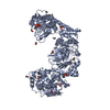
| Structure viewer | Molecule: MolmilJmol/JSmol |
|---|
- Downloads & links
-Download
| PDBx/mmCIF format | 1f3m.cif.gz | 164.4 KB | Display | PDBx/mmCIF format |
|---|---|---|---|---|
| PDB format | pdb1f3m.ent.gz | 128.9 KB | Display | PDB format |
| PDBx/mmJSON format | 1f3m.json.gz | Tree view | PDBx/mmJSON format | |
| Others |  Other downloads Other downloads |
-Validation report
| Arichive directory | https://data.pdbj.org/pub/pdb/validation_reports/f3/1f3mftp://data.pdbj.org/pub/pdb/validation_reports/f3/1f3m | HTTPS FTP |
|---|
-Related structure data
| Similar structure data |
|---|

-Links
 PDBj
PDBj


- Assembly
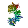
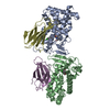
| Deposited unit | 
| ||||||||
|---|---|---|---|---|---|---|---|---|---|
| 1 |
| ||||||||
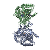
| Unit cell |
|
-Components
| #1: Protein | Mass: 9180.215 Da / Num. of mol.: 2 / Fragment: PAK1 AUTOREGULATORY DOMAIN (70-149) Source method: isolated from a genetically manipulated source Source: (gene. exp.) Homo sapiens (human) / Plasmid: PGEX2N / Production host:  References: UniProt: Q13153, Transferases; Transferring phosphorus-containing groups; Phosphotransferases with an alcohol group as acceptor #2: Protein | Mass: 33273.234 Da / Num. of mol.: 2 / Fragment: KINASE DOMAIN (249-545) / Mutation: K299R Source method: isolated from a genetically manipulated source Source: (gene. exp.) Homo sapiens (human) / Plasmid: PGEX2N / Production host: References: UniProt: Q13153, Transferases; Transferring phosphorus-containing groups; Phosphotransferases with an alcohol group as acceptor #3: Chemical | ChemComp-IOD /   Mass: 126.904 Da / Num. of mol.: 28 / Source method: obtained synthetically / Formula: I Mass: 126.904 Da / Num. of mol.: 28 / Source method: obtained synthetically / Formula: I#4: Water | ChemComp-HOH / |  Mass: 18.015 Da / Num. of mol.: 582 / Source method: isolated from a natural source / Formula: H2O Mass: 18.015 Da / Num. of mol.: 582 / Source method: isolated from a natural source / Formula: H2O |
|---|
-Experimental details
-Experiment
| Experiment | Method: X-RAY DIFFRACTION / Number of used crystals: 1 |
|---|
- Sample preparation
| Crystal | Density Matthews: 3.88 Å3/Da / Density % sol: 68.33 % | |||||||||||||||||||||||||||||||||||||||||||||||||||||||||||||||
|---|---|---|---|---|---|---|---|---|---|---|---|---|---|---|---|---|---|---|---|---|---|---|---|---|---|---|---|---|---|---|---|---|---|---|---|---|---|---|---|---|---|---|---|---|---|---|---|---|---|---|---|---|---|---|---|---|---|---|---|---|---|---|---|---|
| Crystal grow | Temperature: 285 K / Method: vapor diffusion, hanging drop / pH: 6.5 Details: Ammonium Sulfate, pH 6.5, VAPOR DIFFUSION, HANGING DROP, temperature 285K | |||||||||||||||||||||||||||||||||||||||||||||||||||||||||||||||
| Crystal grow | *PLUS Temperature: 12 ℃ / pH: 8 | |||||||||||||||||||||||||||||||||||||||||||||||||||||||||||||||
| Components of the solutions | *PLUS
|
-Data collection
| Diffraction | Mean temperature: 113 K |
|---|---|
| Diffraction source | Source: SYNCHROTRON / Site: APS  / Beamline: 14-BM-C / Wavelength: 1 / Beamline: 14-BM-C / Wavelength: 1 |
| Detector | Type: ADSC QUANTUM 4 / Detector: CCD / Date: Aug 15, 1999 |
| Radiation | Protocol: SINGLE WAVELENGTH / Monochromatic (M) / Laue (L): M / Scattering type: x-ray |
| Radiation wavelength | Wavelength: 1 Å / Relative weight: 1 |
| Reflection | Resolution: 2.3→34.08 Å / Num. all: 56156 / Num. obs: 56156 / % possible obs: 97.9 % / Observed criterion σ(F): -3 / Observed criterion σ(I): -3 / Redundancy: 3 % / Biso Wilson estimate: 32.6 Å2 / Rmerge(I) obs: 0.069 / Net I/σ(I): 15.4 |
| Reflection shell | Resolution: 2.3→2.37 Å / Redundancy: 3 % / Rmerge(I) obs: 0.349 / % possible all: 97.4 |
| Reflection | *PLUS Num. measured all: 160255 |
- Processing
| Software |
| ||||||||||||||||||||
|---|---|---|---|---|---|---|---|---|---|---|---|---|---|---|---|---|---|---|---|---|---|
| Refinement | Resolution: 2.3→34.08 Å / σ(F): 2 / σ(I): 2
| ||||||||||||||||||||
| Refinement step | Cycle: LAST / Resolution: 2.3→34.08 Å
| ||||||||||||||||||||
| Refine LS restraints |
| ||||||||||||||||||||
| Software | *PLUS Name: CNS / Version: 0.9 / Classification: refinement | ||||||||||||||||||||
| Refinement | *PLUS Highest resolution: 2.3 Å / σ(F): 2 / % reflection Rfree: 5 % / Rfactor obs: 0.237 | ||||||||||||||||||||
| Solvent computation | *PLUS | ||||||||||||||||||||
| Displacement parameters | *PLUS |
