 Movie
Movie Controller

[English] 日本語
 Yorodumi
- PDB-7ehh: Crystal structure of alpha-glucosidase from Weissella cibaria BKK... -
+ Open data

- Basic information
| Entry | Database: PDB / ID: 7ehh | ||||||
|---|---|---|---|---|---|---|---|
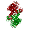
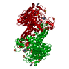
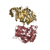
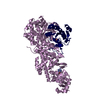
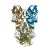
| Title | Crystal structure of alpha-glucosidase from Weissella cibaria BKK1 in complex with maltose | ||||||
 Components Components | alpha-glucosidase | ||||||
 Keywords Keywords | HYDROLASE / glycoside hydrolase / maltooligosaccharides / carbohydrate metabolism / CARBOHYDRATE | ||||||
| Function / homology | Immunoglobulins / Immunoglobulin-like / Sandwich / Mainly Beta Function and homology information Function and homology information | ||||||
| Biological species |  Weissella cibaria (bacteria) Weissella cibaria (bacteria) | ||||||
| Method |  X-RAY DIFFRACTION / SYNCHROTRON / MOLECULAR REPLACEMENT / Resolution: 2 Å X-RAY DIFFRACTION / SYNCHROTRON / MOLECULAR REPLACEMENT / Resolution: 2 Å | ||||||
 Authors Authors | Krusong, K. / Wangpaiboon, K. / Kim, S. / Mori, T. / Hakoshima, T. | ||||||
| Funding support |  Thailand, 1items Thailand, 1items
| ||||||
 Citation Citation | Journal: Acta Crystallogr D Struct Biol / Year: 2021 Title: A GH13 alpha-glucosidase from Weissella cibaria uncommonly acts on short-chain maltooligosaccharides. Authors: Wangpaiboon, K. / Laohawuttichai, P. / Kim, S.Y. / Mori, T. / Nakapong, S. / Pichyangkura, R. / Pongsawasdi, P. / Hakoshima, T. / Krusong, K. | ||||||
| History |
|
- Structure visualization
| Structure viewer | Molecule: MolmilJmol/JSmol |
|---|
- Downloads & links
-Download
| PDBx/mmCIF format | 7ehh.cif.gz | 285.2 KB | Display | PDBx/mmCIF format |
|---|---|---|---|---|
| PDB format | pdb7ehh.ent.gz | 216.7 KB | Display | PDB format |
| PDBx/mmJSON format | 7ehh.json.gz | Tree view | PDBx/mmJSON format | |
| Others |  Other downloads Other downloads |
-Validation report
| Summary document | 7ehh_validation.pdf.gz | 869.5 KB | Display | wwPDB validaton report |
|---|---|---|---|---|
| Full document | 7ehh_full_validation.pdf.gz | 874.2 KB | Display | |
| Data in XML | 7ehh_validation.xml.gz | 27.5 KB | Display | |
| Data in CIF | 7ehh_validation.cif.gz | 41.2 KB | Display | |
| Arichive directory | https://data.pdbj.org/pub/pdb/validation_reports/eh/7ehhftp://data.pdbj.org/pub/pdb/validation_reports/eh/7ehh | HTTPS FTP |
-Related structure data
| Related structure data | 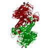 7d9bSC 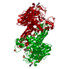 7d9cC 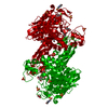 7dcgC 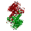 7dchC 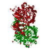 7ehiC S: Starting model for refinement C: citing same article ( |
|---|---|
| Similar structure data |


-Links
 PDBj
PDBj- Assembly
| Deposited unit | 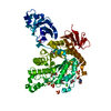
| ||||||||||||
|---|---|---|---|---|---|---|---|---|---|---|---|---|---|
| 1 | 
| ||||||||||||
| Unit cell |
| ||||||||||||
| Components on special symmetry positions |
|
-Components
-Protein / Sugars , 2 types, 2 molecules A
| #1: Protein | Mass: 68398.617 Da / Num. of mol.: 1 Source method: isolated from a genetically manipulated source Source: (gene. exp.) Weissella cibaria (bacteria) / Production host: |
|---|---|
| #2: Polysaccharide | alpha-D-glucopyranose-(1-4)-alpha-D-glucopyranose / alpha-maltose Source method: isolated from a genetically manipulated source |
-Non-polymers , 5 types, 441 molecules 


| #3: Chemical |  Mass: 195.237 Da / Num. of mol.: 2 Mass: 195.237 Da / Num. of mol.: 2Source method: isolated from a genetically manipulated source Formula: C6H13NO4S / Comment: pH buffer*YM #4: Chemical |  Mass: 92.094 Da / Num. of mol.: 3 / Source method: obtained synthetically / Formula: C3H8O3 Mass: 92.094 Da / Num. of mol.: 3 / Source method: obtained synthetically / Formula: C3H8O3#5: Chemical | ChemComp-CA / |  Mass: 40.078 Da / Num. of mol.: 1 / Source method: obtained synthetically / Formula: Ca Mass: 40.078 Da / Num. of mol.: 1 / Source method: obtained synthetically / Formula: Ca#6: Chemical | ChemComp-SO4 /  Mass: 96.063 Da / Num. of mol.: 6 / Source method: obtained synthetically / Formula: SO4 Mass: 96.063 Da / Num. of mol.: 6 / Source method: obtained synthetically / Formula: SO4#7: Water | ChemComp-HOH / | Mass: 18.015 Da / Num. of mol.: 429 / Source method: isolated from a natural source / Formula: H2O |
|---|
-Details
| Has ligand of interest | Y |
|---|---|
| Sequence details | THE SEQUENCE OF THIS PROTEIN WAS NOT AVAILABLE AT THE UNIPROT KNOWLEDGEBASE DATABASE (UNIPROTKB) AT ...THE SEQUENCE OF THIS PROTEIN WAS NOT AVAILABLE AT THE UNIPROT KNOWLEDGEB |
-Experimental details
-Experiment
| Experiment | Method: X-RAY DIFFRACTION / Number of used crystals: 1 |
|---|
- Sample preparation
| Crystal | Density Matthews: 2.64 Å3/Da / Density % sol: 53.45 % |
|---|---|
| Crystal grow | Temperature: 293 K / Method: vapor diffusion / Details: Ammonium sulfate, dioxane, MES |
-Data collection
| Diffraction | Mean temperature: 110 K / Serial crystal experiment: N |
|---|---|
| Diffraction source | Source: SYNCHROTRON / Site: NSRRC  / Beamline: BL13B1 / Wavelength: 1 Å / Beamline: BL13B1 / Wavelength: 1 Å |
| Detector | Type: ADSC QUANTUM 315 / Detector: CCD / Date: Mar 6, 2019 |
| Radiation | Protocol: SINGLE WAVELENGTH / Monochromatic (M) / Laue (L): M / Scattering type: x-ray |
| Radiation wavelength | Wavelength: 1 Å / Relative weight: 1 |
| Reflection | Resolution: 1.8→50 Å / Num. obs: 67707 / % possible obs: 99.6 % / Redundancy: 6.2 % / Biso Wilson estimate: 19.72 Å2 / Rpim(I) all: 0.089 / Rrim(I) all: 0.229 / Net I/σ(I): 19.51 |
| Reflection shell | Resolution: 1.8→1.86 Å / Num. unique obs: 6686 / CC1/2: 0.249 |
- Processing
| Software |
| ||||||||||||||||||||||||||||||||||||||||||||||||||||||||||||||||||||||||||||||||||||||||||||||||||||||||||||||||||||||||||||||
|---|---|---|---|---|---|---|---|---|---|---|---|---|---|---|---|---|---|---|---|---|---|---|---|---|---|---|---|---|---|---|---|---|---|---|---|---|---|---|---|---|---|---|---|---|---|---|---|---|---|---|---|---|---|---|---|---|---|---|---|---|---|---|---|---|---|---|---|---|---|---|---|---|---|---|---|---|---|---|---|---|---|---|---|---|---|---|---|---|---|---|---|---|---|---|---|---|---|---|---|---|---|---|---|---|---|---|---|---|---|---|---|---|---|---|---|---|---|---|---|---|---|---|---|---|---|---|---|
| Refinement | Method to determine structure: MOLECULAR REPLACEMENT Starting model: 7D9B Resolution: 2→32.18 Å / SU ML: 0.1737 / Cross valid method: FREE R-VALUE / σ(F): 1.38 / Phase error: 18.175 Stereochemistry target values: GeoStd + Monomer Library + CDL v1.2
| ||||||||||||||||||||||||||||||||||||||||||||||||||||||||||||||||||||||||||||||||||||||||||||||||||||||||||||||||||||||||||||||
| Solvent computation | Shrinkage radii: 0.9 Å / VDW probe radii: 1.11 Å / Solvent model: FLAT BULK SOLVENT MODEL | ||||||||||||||||||||||||||||||||||||||||||||||||||||||||||||||||||||||||||||||||||||||||||||||||||||||||||||||||||||||||||||||
| Displacement parameters | Biso mean: 23.56 Å2 | ||||||||||||||||||||||||||||||||||||||||||||||||||||||||||||||||||||||||||||||||||||||||||||||||||||||||||||||||||||||||||||||
| Refinement step | Cycle: LAST / Resolution: 2→32.18 Å
| ||||||||||||||||||||||||||||||||||||||||||||||||||||||||||||||||||||||||||||||||||||||||||||||||||||||||||||||||||||||||||||||
| Refine LS restraints |
| ||||||||||||||||||||||||||||||||||||||||||||||||||||||||||||||||||||||||||||||||||||||||||||||||||||||||||||||||||||||||||||||
| LS refinement shell |
|
