Resolution: 3.1→44.8428 Å / Cor.coef. Fo:Fc: 0.909 / Cor.coef. Fo:Fc free: 0.893 / SU B: 16.647 / SU ML: 0.294 / Cross valid method: FREE R-VALUE / ESU R Free: 0.415 Details: Hydrogens have been added in their riding positions
Rfactor
Num. reflection
% reflection
Rfree
0.2101
294
4.946 %
Rwork
0.1813
5650
-
all
0.183
-
-
obs
-
5944
100 %
Solvent computation
Ion probe radii: 0.8 Å / Shrinkage radii: 0.8 Å / VDW probe radii: 1.2 Å / Solvent model: MASK BULK SOLVENT
Displacement parameters
Biso mean: 32.018 Å2
Baniso -1
Baniso -2
Baniso -3
1-
0.056 Å2
0.028 Å2
0 Å2
2-
-
0.056 Å2
-0 Å2
3-
-
-
-0.182 Å2
Refinement step
Cycle: LAST / Resolution: 3.1→44.8428 Å
Protein
Nucleic acid
Ligand
Solvent
Total
Num. atoms
2211
0
0
5
2216
Refine LS restraints
Refine-ID
Type
Dev ideal
Dev ideal target
Number
X-RAY DIFFRACTION
r_bond_refined_d
0.008
0.013
2267
X-RAY DIFFRACTION
r_bond_other_d
0.001
0.017
2165
X-RAY DIFFRACTION
r_angle_refined_deg
1.589
1.641
3062
X-RAY DIFFRACTION
r_angle_other_deg
1.209
1.582
4976
X-RAY DIFFRACTION
r_dihedral_angle_1_deg
7.887
5
277
X-RAY DIFFRACTION
r_dihedral_angle_2_deg
31.463
21.172
128
X-RAY DIFFRACTION
r_dihedral_angle_3_deg
16.956
15
399
X-RAY DIFFRACTION
r_dihedral_angle_4_deg
15.355
15
20
X-RAY DIFFRACTION
r_chiral_restr
0.062
0.2
284
X-RAY DIFFRACTION
r_gen_planes_refined
0.006
0.02
2557
X-RAY DIFFRACTION
r_gen_planes_other
0.001
0.02
539
X-RAY DIFFRACTION
r_nbd_refined
0.225
0.2
550
X-RAY DIFFRACTION
r_symmetry_nbd_other
0.2
0.2
2096
X-RAY DIFFRACTION
r_nbtor_refined
0.169
0.2
1082
X-RAY DIFFRACTION
r_symmetry_nbtor_other
0.075
0.2
1053
X-RAY DIFFRACTION
r_xyhbond_nbd_refined
0.186
0.2
57
X-RAY DIFFRACTION
r_symmetry_xyhbond_nbd_other
0.09
0.2
1
X-RAY DIFFRACTION
r_symmetry_nbd_refined
0.162
0.2
22
X-RAY DIFFRACTION
r_nbd_other
0.219
0.2
79
X-RAY DIFFRACTION
r_symmetry_xyhbond_nbd_refined
0.322
0.2
3
X-RAY DIFFRACTION
r_xyhbond_nbd_other
0.015
0.2
1
X-RAY DIFFRACTION
r_mcbond_it
0.081
3.43
1111
X-RAY DIFFRACTION
r_mcbond_other
0.081
3.43
1110
X-RAY DIFFRACTION
r_mcangle_it
0.157
5.143
1387
X-RAY DIFFRACTION
r_mcangle_other
0.157
5.144
1388
X-RAY DIFFRACTION
r_scbond_it
0.012
3.436
1156
X-RAY DIFFRACTION
r_scbond_other
0.012
3.437
1157
X-RAY DIFFRACTION
r_scangle_it
0.054
5.154
1675
X-RAY DIFFRACTION
r_scangle_other
0.054
5.155
1676
X-RAY DIFFRACTION
r_lrange_it
0.901
39.461
2572
X-RAY DIFFRACTION
r_lrange_other
0.901
39.458
2573
LS refinement shell
Resolution (Å)
Rfactor Rfree
Num. reflection Rfree
Rfactor Rwork
Num. reflection Rwork
Refine-ID
% reflection obs (%)
3.1-3.18
0.233
28
0.223
409
X-RAY DIFFRACTION
100
3.18-3.267
0.23
21
0.22
408
X-RAY DIFFRACTION
100
3.267-3.361
0.229
25
0.214
377
X-RAY DIFFRACTION
100
3.361-3.464
0.23
22
0.196
404
X-RAY DIFFRACTION
100
3.464-3.577
0.233
15
0.193
353
X-RAY DIFFRACTION
100
3.577-3.702
0.16
13
0.18
396
X-RAY DIFFRACTION
100
3.702-3.84
0.27
16
0.184
314
X-RAY DIFFRACTION
100
3.84-3.996
0.096
21
0.187
347
X-RAY DIFFRACTION
100
3.996-4.172
0.22
18
0.161
302
X-RAY DIFFRACTION
100
4.172-4.374
0.24
14
0.167
311
X-RAY DIFFRACTION
100
4.374-4.608
0.225
4
0.161
306
X-RAY DIFFRACTION
100
4.608-4.884
0.184
15
0.169
269
X-RAY DIFFRACTION
100
4.884-5.217
0.129
10
0.177
277
X-RAY DIFFRACTION
100
5.217-5.629
0.203
13
0.201
234
X-RAY DIFFRACTION
100
5.629-6.156
0.29
20
0.186
210
X-RAY DIFFRACTION
100
6.156-6.867
0.278
19
0.181
199
X-RAY DIFFRACTION
100
6.867-7.898
0.109
1
0.143
182
X-RAY DIFFRACTION
100
7.898-9.599
0.168
14
0.124
148
X-RAY DIFFRACTION
100
9.599-13.275
0.317
2
0.139
131
X-RAY DIFFRACTION
100
+
About Yorodumi
-
News
-
Feb 9, 2022. New format data for meta-information of EMDB entries
New format data for meta-information of EMDB entries
Version 3 of the EMDB header file is now the official format.
The previous official version 1.9 will be removed from the archive.
In the structure databanks used in Yorodumi, some data are registered as the other names, "COVID-19 virus" and "2019-nCoV". Here are the details of the virus and the list of structure data.
Jan 31, 2019. EMDB accession codes are about to change! (news from PDBe EMDB page)
EMDB accession codes are about to change! (news from PDBe EMDB page)
The allocation of 4 digits for EMDB accession codes will soon come to an end. Whilst these codes will remain in use, new EMDB accession codes will include an additional digit and will expand incrementally as the available range of codes is exhausted. The current 4-digit format prefixed with “EMD-” (i.e. EMD-XXXX) will advance to a 5-digit format (i.e. EMD-XXXXX), and so on. It is currently estimated that the 4-digit codes will be depleted around Spring 2019, at which point the 5-digit format will come into force.
The EM Navigator/Yorodumi systems omit the EMD- prefix.
Related info.:Q: What is EMD? / ID/Accession-code notation in Yorodumi/EM Navigator
Yorodumi is a browser for structure data from EMDB, PDB, SASBDB, etc.
This page is also the successor to EM Navigator detail page, and also detail information page/front-end page for Omokage search.
The word "yorodu" (or yorozu) is an old Japanese word meaning "ten thousand". "mi" (miru) is to see.
Related info.:EMDB / PDB / SASBDB / Comparison of 3 databanks / Yorodumi Search / Aug 31, 2016. New EM Navigator & Yorodumi / Yorodumi Papers / Jmol/JSmol / Function and homology information / Changes in new EM Navigator and Yorodumi
 Movie
Movie Controller


 Open data
 Basic information
 Components
 Keywords
 Function and homology information
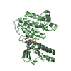
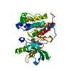
 Homo sapiens (human)
 X-RAY DIFFRACTION /
  Authors
 Citation
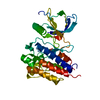
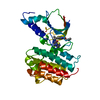
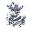
 Structure visualization
 Downloads & links
 Other downloads


 PDBj
PDBj


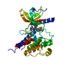
 Assembly
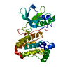

 Mass: 18.015 Da / Num. of mol.: 5 / Source method: isolated from a natural source / Formula: H2O
 Sample preparation
 Processing