| Entry | Database: PDB / ID: 7ed1
|
|---|
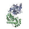
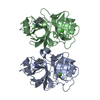
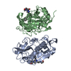
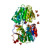
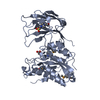
| Title | Transferase from Mycobacterium smegmatis |
|---|
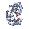
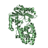
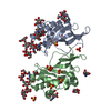
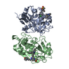
 Components Components | 4'-phosphopantetheinyl transferase |
|---|
 Keywords Keywords | TRANSFERASE / phosphopantetheine transferase |
|---|
| Function / homology | COENZYME A / 4'-phosphopantetheinyl transferase Function and homology information Function and homology information |
|---|
| Biological species |  Mycolicibacterium smegmatis (bacteria) Mycolicibacterium smegmatis (bacteria) |
|---|
| Method |  X-RAY DIFFRACTION / SYNCHROTRON / MOLECULAR REPLACEMENT / Resolution: 1.65 Å X-RAY DIFFRACTION / SYNCHROTRON / MOLECULAR REPLACEMENT / Resolution: 1.65 Å |
|---|
 Authors Authors | Kim, D.-G. / Lee, B.-J. / Lee, S.J. |
|---|
| Funding support |  Korea, Republic Of, 3items Korea, Republic Of, 3items | Organization | Grant number | Country |
|---|
| National Research Foundation (NRF, Korea) | NRF-2018R1A2A1A19018526 | Korea, Republic Of | | National Research Foundation (NRF, Korea) | NRF-2018R1A5A2024425 | Korea, Republic Of | | National Research Foundation (NRF, Korea) | 2020R1F1A1075828 | Korea, Republic Of |
|
|---|
 Citation Citation | Journal: To Be Published
Title: Transferase from Mycobacterium smegmatis
Authors: Kim, D.-G. / Lee, B.-J. / Lee, S.J. |
|---|
| History | | Deposition | Mar 15, 2021 | Deposition site: PDBJ / Processing site: PDBJ |
|---|
| Revision 1.0 | Mar 16, 2022 | Provider: repository / Type: Initial release |
|---|
| Revision 1.1 | May 29, 2024 | Group: Data collection / Category: chem_comp_atom / chem_comp_bond |
|---|
| Revision 2.0 | Aug 14, 2024 | Group: Advisory / Atomic model ...Advisory / Atomic model / Author supporting evidence / Data collection / Database references / Derived calculations / Non-polymer description / Polymer sequence / Refinement description / Source and taxonomy / Structure summary
Category: atom_site / atom_type ...atom_site / atom_type / chem_comp / chem_comp_atom / chem_comp_bond / citation / entity / entity_poly / entity_poly_seq / entity_src_gen / pdbx_contact_author / pdbx_entity_instance_feature / pdbx_entity_nonpoly / pdbx_nonpoly_scheme / pdbx_poly_seq_scheme / pdbx_struct_assembly / pdbx_struct_conn_angle / pdbx_struct_sheet_hbond / pdbx_unobs_or_zero_occ_residues / refine / refine_hist / refine_ls_restr / refine_ls_shell / struct / struct_conf / struct_conn / struct_ref / struct_ref_seq / struct_ref_seq_dif / struct_sheet_range
Item: _atom_site.auth_atom_id / _atom_site.auth_comp_id ..._atom_site.auth_atom_id / _atom_site.auth_comp_id / _atom_site.label_atom_id / _atom_site.label_comp_id / _atom_site.label_seq_id / _atom_site.type_symbol / _chem_comp.formula / _chem_comp.formula_weight / _chem_comp.id / _chem_comp.mon_nstd_flag / _chem_comp.name / _chem_comp.type / _citation.title / _entity.formula_weight / _entity.pdbx_description / _entity_poly.pdbx_seq_one_letter_code / _entity_poly.pdbx_seq_one_letter_code_can / _entity_src_gen.gene_src_common_name / _entity_src_gen.pdbx_end_seq_num / _pdbx_entity_instance_feature.auth_comp_id / _pdbx_entity_instance_feature.comp_id / _pdbx_entity_nonpoly.comp_id / _pdbx_entity_nonpoly.name / _pdbx_nonpoly_scheme.auth_mon_id / _pdbx_nonpoly_scheme.mon_id / _pdbx_nonpoly_scheme.pdb_mon_id / _pdbx_struct_assembly.details / _pdbx_struct_sheet_hbond.range_1_label_seq_id / _pdbx_struct_sheet_hbond.range_2_label_seq_id / _refine.B_iso_max / _refine.B_iso_min / _refine.details / _refine.ls_R_factor_R_free / _refine.ls_R_factor_R_work / _refine.ls_R_factor_obs / _refine.pdbx_ls_sigma_F / _refine_hist.cycle_id / _refine_hist.pdbx_B_iso_mean_ligand / _refine_hist.pdbx_B_iso_mean_solvent / _refine_hist.pdbx_number_residues_total / _refine_ls_shell.R_factor_R_free_error / _refine_ls_shell.number_reflns_all / _struct.title / _struct_conf.beg_label_seq_id / _struct_conf.end_label_seq_id / _struct_ref.db_code / _struct_ref.pdbx_align_begin / _struct_ref.pdbx_db_accession / _struct_ref.pdbx_seq_one_letter_code / _struct_ref_seq.db_align_beg / _struct_ref_seq.pdbx_auth_seq_align_beg / _struct_ref_seq.pdbx_db_accession / _struct_ref_seq.seq_align_end / _struct_sheet_range.beg_label_seq_id / _struct_sheet_range.end_label_seq_id
Description: Ligand identity / Details: Ca ions to Mg ions / Provider: author / Type: Coordinate replacement |
|---|
|
|---|
 Movie
Movie Controller


 Open data
 Basic information
 Structure visualization
 Downloads & links
 Other downloads
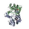
 PDBj
PDBj


 Assembly
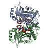

 Mass: 767.534 Da / Num. of mol.: 2 / Source method: obtained synthetically / Formula: C21H36N7O16P3S / Feature type: SUBJECT OF INVESTIGATION
 Mass: 238.278 Da / Num. of mol.: 2 / Source method: obtained synthetically / Formula: C10H22O6 / Feature type: SUBJECT OF INVESTIGATION / Comment: precipitant*YM
 Mass: 24.305 Da / Num. of mol.: 6 / Source method: obtained synthetically / Formula: Mg / Feature type: SUBJECT OF INVESTIGATION
 Mass: 18.015 Da / Num. of mol.: 378 / Source method: isolated from a natural source / Formula: H2O
 Sample preparation
 Processing