 Movie
Movie Controller

[English] 日本語
 Yorodumi
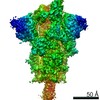
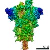
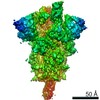
- PDB-7eaz: Cryo-EM structure of SARS-CoV-2 Spike D614G variant, one RBD-up c... -
+ Open data

- Basic information
| Entry | Database: PDB / ID: 7eaz | |||||||||||||||||||||||||||
|---|---|---|---|---|---|---|---|---|---|---|---|---|---|---|---|---|---|---|---|---|---|---|---|---|---|---|---|---|
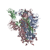
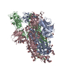
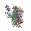
| Title | Cryo-EM structure of SARS-CoV-2 Spike D614G variant, one RBD-up conformation 1 | |||||||||||||||||||||||||||
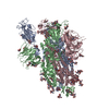
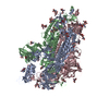
 Components Components | Spike glycoprotein | |||||||||||||||||||||||||||
 Keywords Keywords | VIRAL PROTEIN / SARS-CoV-2 / Spike protein | |||||||||||||||||||||||||||
| Function / homology |  Function and homology information Function and homology informationsymbiont-mediated disruption of host tissue / Maturation of spike protein / Translation of Structural Proteins / Virion Assembly and Release / host cell surface / host extracellular region / symbiont-mediated-mediated suppression of host tetherin activity / Induction of Cell-Cell Fusion / structural constituent of virion / positive regulation of viral entry into host cell ...symbiont-mediated disruption of host tissue / Maturation of spike protein / Translation of Structural Proteins / Virion Assembly and Release / host cell surface / host extracellular region / symbiont-mediated-mediated suppression of host tetherin activity / Induction of Cell-Cell Fusion / structural constituent of virion / positive regulation of viral entry into host cell / membrane fusion / host cell endoplasmic reticulum-Golgi intermediate compartment membrane / Attachment and Entry / entry receptor-mediated virion attachment to host cell / receptor-mediated virion attachment to host cell / host cell surface receptor binding / symbiont-mediated suppression of host innate immune response / endocytosis involved in viral entry into host cell / receptor ligand activity / fusion of virus membrane with host plasma membrane / fusion of virus membrane with host endosome membrane / viral envelope / symbiont entry into host cell / virion attachment to host cell / host cell plasma membrane / SARS-CoV-2 activates/modulates innate and adaptive immune responses / virion membrane / membrane / identical protein binding / plasma membrane Similarity search - Function | |||||||||||||||||||||||||||
| Biological species |   Severe acute respiratory syndrome coronavirus 2 Severe acute respiratory syndrome coronavirus 2 | |||||||||||||||||||||||||||
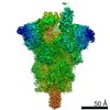
| Method | ELECTRON MICROSCOPY / single particle reconstruction / cryo EM / Resolution: 3.5 Å | |||||||||||||||||||||||||||
 Authors Authors | Yang, T.J. / Yu, P.Y. / Chang, Y.C. / Hsu, S.T.D. | |||||||||||||||||||||||||||
| Funding support |  Taiwan, 4items Taiwan, 4items
| |||||||||||||||||||||||||||
 Citation Citation | Journal: J Biol Chem / Year: 2021 Title: D614G mutation in the SARS-CoV-2 spike protein enhances viral fitness by desensitizing it to temperature-dependent denaturation. Authors: Tzu-Jing Yang / Pei-Yu Yu / Yuan-Chih Chang / Shang-Te Danny Hsu / Abstract: The D614G mutation in the spike protein of SARS-CoV-2 alters the fitness of the virus, leading to the dominant form observed in the COVID-19 pandemic. However, the molecular basis of the mechanism by ...The D614G mutation in the spike protein of SARS-CoV-2 alters the fitness of the virus, leading to the dominant form observed in the COVID-19 pandemic. However, the molecular basis of the mechanism by which this mutation enhances fitness is not clear. Here we demonstrated by cryo-electron microscopy that the D614G mutation resulted in increased propensity of multiple receptor-binding domains (RBDs) in an upward conformation poised for host receptor binding. Multiple substates within the one RBD-up or two RBD-up conformational space were determined. According to negative staining electron microscopy, differential scanning calorimetry, and differential scanning fluorimetry, the most significant impact of the mutation lies in its ability to eliminate the unusual cold-induced unfolding characteristics and to significantly increase the thermal stability under physiological pH. The D614G spike variant also exhibited exceptional long-term stability when stored at 37 °C for up to 2 months. Our findings shed light on how the D614G mutation enhances the infectivity of SARS-CoV-2 through a stabilizing mutation and suggest an approach for better design of spike protein-based conjugates for vaccine development. | |||||||||||||||||||||||||||
| History |
|
- Structure visualization
| Movie |
Movie viewer |
|---|---|
| Structure viewer | Molecule: MolmilJmol/JSmol |
- Downloads & links
-Download
| PDBx/mmCIF format | 7eaz.cif.gz | 566.1 KB | Display | PDBx/mmCIF format |
|---|---|---|---|---|
| PDB format | pdb7eaz.ent.gz | 462.3 KB | Display | PDB format |
| PDBx/mmJSON format | 7eaz.json.gz | Tree view | PDBx/mmJSON format | |
| Others |  Other downloads Other downloads |
-Validation report
| Arichive directory | https://data.pdbj.org/pub/pdb/validation_reports/ea/7eazftp://data.pdbj.org/pub/pdb/validation_reports/ea/7eaz | HTTPS FTP |
|---|
-Related structure data
| Related structure data | 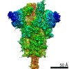 31047MC  7eb0C 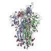 7eb3C 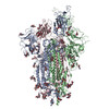 7eb4C 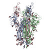 7eb5C M: map data used to model this data C: citing same article ( |
|---|---|
| Similar structure data |


-Links
 PDBj
PDBj


- Assembly
| Deposited unit | 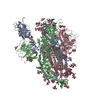
|
|---|---|
| 1 |
|
-Components
| #1: Protein | Mass: 142178.297 Da / Num. of mol.: 3 Source method: isolated from a genetically manipulated source Source: (gene. exp.) Severe acute respiratory syndrome coronavirus 2Gene: S, 2 / Production host:  Homo sapiens (human) / References: UniProt: P0DTC2 Homo sapiens (human) / References: UniProt: P0DTC2#2: Polysaccharide | Source method: isolated from a genetically manipulated source #3: Polysaccharide | 2-acetamido-2-deoxy-beta-D-glucopyranose-(1-4)-2-acetamido-2-deoxy-beta-D-glucopyranose Source method: isolated from a genetically manipulated source #4: Sugar | ChemComp-NAG /   Type: D-saccharide, beta linking / Mass: 221.208 Da / Num. of mol.: 16 / Source method: obtained synthetically / Formula: C8H15NO6 Type: D-saccharide, beta linking / Mass: 221.208 Da / Num. of mol.: 16 / Source method: obtained synthetically / Formula: C8H15NO6Has ligand of interest | N | Has protein modification | Y | Sequence details | The residues 682-685 (GSAS) and residues 986-987 (PP) are designed for protein stabilization. | |
|---|
-Experimental details
-Experiment
| Experiment | Method: ELECTRON MICROSCOPY |
|---|---|
| EM experiment | Aggregation state: PARTICLE / 3D reconstruction method: single particle reconstruction |
- Sample preparation
| Component | Name: SARS-CoV-2 spike glycoprotein / Type: ORGANELLE OR CELLULAR COMPONENT / Details: D614G variant, one RBD-up conformation 1 / Entity ID: #1 / Source: RECOMBINANT | ||||||||||||||||||||
|---|---|---|---|---|---|---|---|---|---|---|---|---|---|---|---|---|---|---|---|---|---|
| Molecular weight | Value: 0.54 MDa / Experimental value: YES | ||||||||||||||||||||
| Source (natural) | Organism: Severe acute respiratory syndrome coronavirus 2 | ||||||||||||||||||||
| Source (recombinant) | Organism: Homo sapiens (human) | ||||||||||||||||||||
| Buffer solution | pH: 7.6 | ||||||||||||||||||||
| Buffer component |
| ||||||||||||||||||||
| Specimen | Conc.: 1 mg/ml / Embedding applied: NO / Shadowing applied: NO / Staining applied: NO / Vitrification applied: YES | ||||||||||||||||||||
| Vitrification | Instrument: FEI VITROBOT MARK IV / Cryogen name: ETHANE / Humidity: 100 % / Chamber temperature: 277 K Details: blot for 2.5 seconds before plunging; blot force: 0; waiting time: 30s |
- Electron microscopy imaging
| Experimental equipment |  Model: Titan Krios / Image courtesy: FEI Company |
|---|---|
| Microscopy | Model: FEI TITAN KRIOS |
| Electron gun | Electron source:  FIELD EMISSION GUN / Accelerating voltage: 300 kV / Illumination mode: FLOOD BEAM FIELD EMISSION GUN / Accelerating voltage: 300 kV / Illumination mode: FLOOD BEAM |
| Electron lens | Mode: BRIGHT FIELD / Cs: 2.7 mm |
| Specimen holder | Cryogen: NITROGEN |
| Image recording | Electron dose: 1 e/Å2 / Film or detector model: GATAN K3 (6k x 4k) |
- Processing
| Software | Name: PHENIX / Version: 1.19.1_4122: / Classification: refinement | ||||||||||||||||||||||||
|---|---|---|---|---|---|---|---|---|---|---|---|---|---|---|---|---|---|---|---|---|---|---|---|---|---|
| EM software |
| ||||||||||||||||||||||||
| CTF correction | Type: PHASE FLIPPING AND AMPLITUDE CORRECTION | ||||||||||||||||||||||||
| Symmetry | Point symmetry: C1 (asymmetric) | ||||||||||||||||||||||||
| 3D reconstruction | Resolution: 3.5 Å / Resolution method: FSC 0.143 CUT-OFF / Num. of particles: 104186 / Symmetry type: POINT | ||||||||||||||||||||||||
| Refine LS restraints |
|
