 Movie
Movie Controller

[English] 日本語
 Yorodumi
- PDB-7e7m: Crystal Structure Analysis of the Streptococcus agalactiae Ribose... -
+ Open data

- Basic information
| Entry | Database: PDB / ID: 7e7m | |||||||||
|---|---|---|---|---|---|---|---|---|---|---|
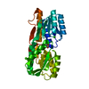
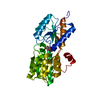
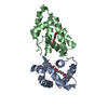
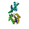
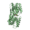
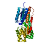
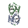
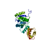
| Title | Crystal Structure Analysis of the Streptococcus agalactiae Ribose Binding Protein RbsB | |||||||||
 Components Components | D-ribose ABC transporter substrate-binding protein | |||||||||
 Keywords Keywords | SUGAR BINDING PROTEIN / Protein-ribose complex | |||||||||
| Function / homology | beta-D-ribopyranose / :  Function and homology information Function and homology information | |||||||||
| Biological species |  Streptococcus agalactiae ZQ0910 (bacteria) Streptococcus agalactiae ZQ0910 (bacteria) | |||||||||
| Method |  X-RAY DIFFRACTION / SYNCHROTRON / MOLECULAR REPLACEMENT / Resolution: 2.85 Å X-RAY DIFFRACTION / SYNCHROTRON / MOLECULAR REPLACEMENT / Resolution: 2.85 Å | |||||||||
 Authors Authors | Pan, L.X. / Wang, B. / Yang, L.Y. / Yang, D.F. | |||||||||
| Funding support |  China, 2items China, 2items
| |||||||||
 Citation Citation | Journal: To be published Title: Crystal Structure Analysis of the Streptococcus agalactiae Ribose Binding Protein RbsB Authors: Pan, L.X. / Wang, B. / Yang, L.Y. / Yang, D.F. | |||||||||
| History |
|
- Structure visualization
| Structure viewer | Molecule: MolmilJmol/JSmol |
|---|
- Downloads & links
-Download
| PDBx/mmCIF format | 7e7m.cif.gz | 214.9 KB | Display | PDBx/mmCIF format |
|---|---|---|---|---|
| PDB format | pdb7e7m.ent.gz | 170.6 KB | Display | PDB format |
| PDBx/mmJSON format | 7e7m.json.gz | Tree view | PDBx/mmJSON format | |
| Others |  Other downloads Other downloads |
-Validation report
| Arichive directory | https://data.pdbj.org/pub/pdb/validation_reports/e7/7e7mftp://data.pdbj.org/pub/pdb/validation_reports/e7/7e7m | HTTPS FTP |
|---|
-Related structure data
| Related structure data |  2ioyS S: Starting model for refinement |
|---|---|
| Similar structure data |

-Links
 PDBj
PDBj- Assembly
| Deposited unit | 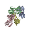
| ||||||||
|---|---|---|---|---|---|---|---|---|---|
| 1 | 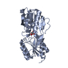
| ||||||||
| 2 | 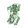
| ||||||||
| 3 | 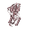
| ||||||||
| 4 | 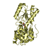
| ||||||||
| Unit cell |
|
-Components
| #1: Protein | Mass: 34124.129 Da / Num. of mol.: 4 Source method: isolated from a genetically manipulated source Source: (gene. exp.) Streptococcus agalactiae ZQ0910 (bacteria)Gene: rbsB / Plasmid: pGEX-6p-1 / Production host: #2: Sugar | ChemComp-RIP / 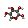  Type: D-saccharide, beta linking / Mass: 150.130 Da / Num. of mol.: 4 / Source method: obtained synthetically / Formula: C5H10O5 / Feature type: SUBJECT OF INVESTIGATION Type: D-saccharide, beta linking / Mass: 150.130 Da / Num. of mol.: 4 / Source method: obtained synthetically / Formula: C5H10O5 / Feature type: SUBJECT OF INVESTIGATION#3: Water | ChemComp-HOH / |  Mass: 18.015 Da / Num. of mol.: 8 / Source method: isolated from a natural source / Formula: H2O Mass: 18.015 Da / Num. of mol.: 8 / Source method: isolated from a natural source / Formula: H2OHas ligand of interest | Y | |
|---|
-Experimental details
-Experiment
| Experiment | Method: X-RAY DIFFRACTION / Number of used crystals: 1 |
|---|
- Sample preparation
| Crystal | Density Matthews: 1.86 Å3/Da / Density % sol: 34.03 % / Description: plate |
|---|---|
| Crystal grow | Temperature: 293 K / Method: evaporation / pH: 7 / Details: jeffamime ED-2001 |
-Data collection
| Diffraction | Mean temperature: 100 K / Serial crystal experiment: N | ||||||||||||||||||||||||
|---|---|---|---|---|---|---|---|---|---|---|---|---|---|---|---|---|---|---|---|---|---|---|---|---|---|
| Diffraction source | Source: SYNCHROTRON / Site: SSRF / Beamline: BL18U1 / Wavelength: 0.97915 Å | ||||||||||||||||||||||||
| Detector | Type: DECTRIS PILATUS3 6M / Detector: PIXEL / Date: Jan 5, 2021 | ||||||||||||||||||||||||
| Radiation | Protocol: SINGLE WAVELENGTH / Monochromatic (M) / Laue (L): M / Scattering type: x-ray | ||||||||||||||||||||||||
| Radiation wavelength | Wavelength: 0.97915 Å / Relative weight: 1 | ||||||||||||||||||||||||
| Reflection | Resolution: 2.85→39.63 Å / Num. obs: 22672 / % possible obs: 94.4 % / Redundancy: 5.4 % / Biso Wilson estimate: 54.12 Å2 / CC1/2: 0.996 / Rmerge(I) obs: 0.122 / Rpim(I) all: 0.055 / Rrim(I) all: 0.134 / Net I/σ(I): 11.7 / Num. measured all: 121350 / Scaling rejects: 7 | ||||||||||||||||||||||||
| Reflection shell | Diffraction-ID: 1
|
- Processing
| Software |
| |||||||||||||||||||||||||||||||||||||||||||||||||||||||||||||||
|---|---|---|---|---|---|---|---|---|---|---|---|---|---|---|---|---|---|---|---|---|---|---|---|---|---|---|---|---|---|---|---|---|---|---|---|---|---|---|---|---|---|---|---|---|---|---|---|---|---|---|---|---|---|---|---|---|---|---|---|---|---|---|---|---|
| Refinement | Method to determine structure: MOLECULAR REPLACEMENT Starting model: 2IOY Resolution: 2.85→38.59 Å / SU ML: 0.36 / Cross valid method: THROUGHOUT / σ(F): 1.35 / Phase error: 27.15 / Stereochemistry target values: ML
| |||||||||||||||||||||||||||||||||||||||||||||||||||||||||||||||
| Solvent computation | Shrinkage radii: 0.9 Å / VDW probe radii: 1.11 Å / Solvent model: FLAT BULK SOLVENT MODEL | |||||||||||||||||||||||||||||||||||||||||||||||||||||||||||||||
| Displacement parameters | Biso max: 190.51 Å2 / Biso mean: 57.5412 Å2 / Biso min: 26.03 Å2 | |||||||||||||||||||||||||||||||||||||||||||||||||||||||||||||||
| Refinement step | Cycle: final / Resolution: 2.85→38.59 Å
| |||||||||||||||||||||||||||||||||||||||||||||||||||||||||||||||
| LS refinement shell | Refine-ID: X-RAY DIFFRACTION / Rfactor Rfree error: 0 / Total num. of bins used: 8
|
