 Movie
Movie Controller

[English] 日本語
 Yorodumi
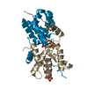
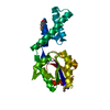
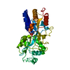
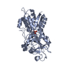
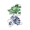
- PDB-2ra6: Crystal Structure of the Possum Milk Whey Lipocalin Trichosurin a... -
+ Open data

- Basic information
| Entry | Database: PDB / ID: 2ra6 | ||||||
|---|---|---|---|---|---|---|---|
| Title | Crystal Structure of the Possum Milk Whey Lipocalin Trichosurin at pH 4.6 with Bound 4-ethylphenol | ||||||
 Components Components | Trichosurin | ||||||
 Keywords Keywords | TRANSPORT PROTEIN / Lipocalin / beta Barrel / Glycoprotein / Milk protein / Secreted / Transport | ||||||
| Function / homology |  Function and homology information Function and homology information | ||||||
| Biological species |  | ||||||
| Method |  X-RAY DIFFRACTION / SYNCHROTRON / MOLECULAR REPLACEMENT / Resolution: 1.5 Å X-RAY DIFFRACTION / SYNCHROTRON / MOLECULAR REPLACEMENT / Resolution: 1.5 Å | ||||||
 Authors Authors | Watson, R.P. | ||||||
 Citation Citation | Journal: Biochem.J. / Year: 2007 Title: Three-dimensional structure and ligand binding properties of trichosurin, a metatherian lipocalin from the milk whey of the common brushtail possum Trichosurus vulpecula Authors: Watson, R.P. / Demmer, J. / Baker, E.N. / Arcus, V.L. | ||||||
| History |
|
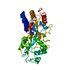
- Structure visualization
| Structure viewer | Molecule: MolmilJmol/JSmol |
|---|
- Downloads & links
-Download
| PDBx/mmCIF format | 2ra6.cif.gz | 138.2 KB | Display | PDBx/mmCIF format |
|---|---|---|---|---|
| PDB format | pdb2ra6.ent.gz | 108.9 KB | Display | PDB format |
| PDBx/mmJSON format | 2ra6.json.gz | Tree view | PDBx/mmJSON format | |
| Others |  Other downloads Other downloads |
-Validation report
| Arichive directory | https://data.pdbj.org/pub/pdb/validation_reports/ra/2ra6ftp://data.pdbj.org/pub/pdb/validation_reports/ra/2ra6 | HTTPS FTP |
|---|
-Related structure data
| Related structure data |  2r73C  2r74SC S: Starting model for refinement C: citing same article ( |
|---|---|
| Similar structure data |

-Links
 PDBj
PDBj


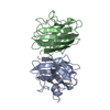
- Assembly
| Deposited unit | 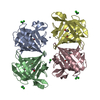
| ||||||||
|---|---|---|---|---|---|---|---|---|---|
| 1 | 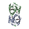
| ||||||||
| 2 | 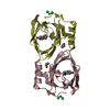
| ||||||||
| Unit cell |
|
-Components
-Protein , 1 types, 4 molecules ABCD
| #1: Protein | Mass: 19709.904 Da / Num. of mol.: 4 / Mutation: G102E Source method: isolated from a genetically manipulated source Source: (gene. exp.) Plasmid: pET23a / Species (production host): Escherichia coli / Production host:  |
|---|
-Non-polymers , 5 types, 407 molecules 

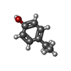


| #2: Chemical | ChemComp-ZN /  Mass: 65.409 Da / Num. of mol.: 6 / Source method: obtained synthetically / Formula: Zn Mass: 65.409 Da / Num. of mol.: 6 / Source method: obtained synthetically / Formula: Zn#3: Chemical | ChemComp-CL /  Mass: 35.453 Da / Num. of mol.: 9 / Source method: obtained synthetically / Formula: Cl Mass: 35.453 Da / Num. of mol.: 9 / Source method: obtained synthetically / Formula: Cl#4: Chemical | ChemComp-ETY /  Mass: 122.164 Da / Num. of mol.: 4 / Source method: obtained synthetically / Formula: C8H10O Mass: 122.164 Da / Num. of mol.: 4 / Source method: obtained synthetically / Formula: C8H10O#5: Chemical | ChemComp-IPA /  Mass: 60.095 Da / Num. of mol.: 8 / Source method: obtained synthetically / Formula: C3H8O Mass: 60.095 Da / Num. of mol.: 8 / Source method: obtained synthetically / Formula: C3H8O#6: Water | ChemComp-HOH / | Mass: 18.015 Da / Num. of mol.: 380 / Source method: isolated from a natural source / Formula: H2O |
|---|
-Details
| Has protein modification | Y |
|---|
-Experimental details
-Experiment
| Experiment | Method: X-RAY DIFFRACTION / Number of used crystals: 1 |
|---|
- Sample preparation
| Crystal | Density Matthews: 2.39 Å3/Da / Density % sol: 48.58 % |
|---|---|
| Crystal grow | Temperature: 291 K / Method: vapor diffusion, hanging drop / pH: 4.6 Details: 100mM Sodium Acetate, 200mM ZnCl2, 8% 2-propanol, pH 4.6, VAPOR DIFFUSION, HANGING DROP, temperature 291K |
-Data collection
| Diffraction | Mean temperature: 113 K |
|---|---|
| Diffraction source | Source: SYNCHROTRON / Site: ESRF  / Beamline: ID14-4 / Wavelength: 0.89 Å / Beamline: ID14-4 / Wavelength: 0.89 Å |
| Detector | Type: ADSC QUANTUM 4 / Detector: CCD / Date: Sep 1, 2003 |
| Radiation | Protocol: SINGLE WAVELENGTH / Monochromatic (M) / Laue (L): M / Scattering type: x-ray |
| Radiation wavelength | Wavelength: 0.89 Å / Relative weight: 1 |
| Reflection | Resolution: 1.5→30 Å / Num. all: 118308 / Num. obs: 117706 / % possible obs: 99.5 % / Observed criterion σ(F): 1 / Observed criterion σ(I): 1 / Rmerge(I) obs: 0.072 / Net I/σ(I): 11.6 |
| Reflection shell | Resolution: 1.5→1.6 Å / Rmerge(I) obs: 0.289 / Mean I/σ(I) obs: 4.3 / % possible all: 96 |
- Processing
| Software |
| |||||||||||||||||||||||||
|---|---|---|---|---|---|---|---|---|---|---|---|---|---|---|---|---|---|---|---|---|---|---|---|---|---|---|
| Refinement | Method to determine structure: MOLECULAR REPLACEMENT Starting model: PDB entry 2R74 Resolution: 1.5→20 Å / σ(F): 0 / σ(I): 0 / Stereochemistry target values: Engh & Huber
| |||||||||||||||||||||||||
| Refinement step | Cycle: LAST / Resolution: 1.5→20 Å
| |||||||||||||||||||||||||
| Refine LS restraints |
|
