 Movie
Movie Controller

[English] 日本語
 Yorodumi
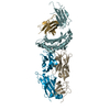
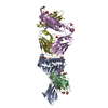
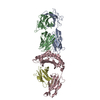
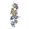
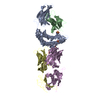
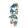
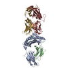
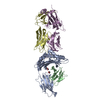
- PDB-7e72: Crystal structure of Tie2-agonistic antibody in complex with huma... -
+ Open data

- Basic information
| Entry | Database: PDB / ID: 7.0E+72 | |||||||||
|---|---|---|---|---|---|---|---|---|---|---|
| Title | Crystal structure of Tie2-agonistic antibody in complex with human Tie2 Fn2-3 | |||||||||
 Components Components |
| |||||||||
 Keywords Keywords | MEMBRANE PROTEIN / antigen-antibody complex / agonistic antibody / angiogenesis / vascular stabilization / receptor tyrosine kinase / Tie2 | |||||||||
| Function / homology |  Function and homology information Function and homology informationTie signaling pathway / glomerulus vasculature development / regulation of endothelial cell apoptotic process / regulation of establishment or maintenance of cell polarity / regulation of vascular permeability / heart trabecula formation / definitive hemopoiesis / sprouting angiogenesis / endothelial cell proliferation / positive regulation of Rho protein signal transduction ...Tie signaling pathway / glomerulus vasculature development / regulation of endothelial cell apoptotic process / regulation of establishment or maintenance of cell polarity / regulation of vascular permeability / heart trabecula formation / definitive hemopoiesis / sprouting angiogenesis / endothelial cell proliferation / positive regulation of Rho protein signal transduction / microvillus / positive regulation of Rac protein signal transduction / positive regulation of intracellular signal transduction / positive regulation of focal adhesion assembly / negative regulation of endothelial cell apoptotic process / Tie2 Signaling / positive regulation of endothelial cell proliferation / transmembrane receptor protein tyrosine kinase activity / positive regulation of endothelial cell migration / substrate adhesion-dependent cell spreading / cell surface receptor protein tyrosine kinase signaling pathway / basal plasma membrane / negative regulation of angiogenesis / cellular response to mechanical stimulus / receptor protein-tyrosine kinase / negative regulation of inflammatory response / positive regulation of angiogenesis / cell-cell junction / cell-cell signaling / signaling receptor activity / heart development / RAF/MAP kinase cascade / angiogenesis / basolateral plasma membrane / cell surface receptor signaling pathway / positive regulation of ERK1 and ERK2 cascade / positive regulation of phosphatidylinositol 3-kinase/protein kinase B signal transduction / protein kinase activity / receptor complex / positive regulation of MAPK cascade / ciliary basal body / apical plasma membrane / membrane raft / focal adhesion / centrosome / negative regulation of apoptotic process / cell surface / extracellular region / ATP binding / identical protein binding / plasma membrane / cytoplasm Similarity search - Function | |||||||||
| Biological species |  Homo sapiens (human) Homo sapiens (human) | |||||||||
| Method |  X-RAY DIFFRACTION / SYNCHROTRON / MOLECULAR REPLACEMENT / Resolution: 2.094 Å X-RAY DIFFRACTION / SYNCHROTRON / MOLECULAR REPLACEMENT / Resolution: 2.094 Å | |||||||||
 Authors Authors | Kim, H.M. / Jo, G.H. / Hong, H.J. / Han, A. | |||||||||
| Funding support |  Korea, Republic Of, 2items Korea, Republic Of, 2items
| |||||||||
 Citation Citation | Journal: Nat Commun / Year: 2021 Title: Structural insights into the clustering and activation of Tie2 receptor mediated by Tie2 agonistic antibody. Authors: Jo, G. / Bae, J. / Hong, H.J. / Han, A.R. / Kim, D.K. / Hong, S.P. / Kim, J.A. / Lee, S. / Koh, G.Y. / Kim, H.M. | |||||||||
| History |
|
- Structure visualization
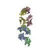
| Structure viewer | Molecule: MolmilJmol/JSmol |
|---|
- Downloads & links
-Download
| PDBx/mmCIF format | 7e72.cif.gz | 518.3 KB | Display | PDBx/mmCIF format |
|---|---|---|---|---|
| PDB format | pdb7e72.ent.gz | 428.6 KB | Display | PDB format |
| PDBx/mmJSON format | 7e72.json.gz | Tree view | PDBx/mmJSON format | |
| Others |  Other downloads Other downloads |
-Validation report
| Summary document | 7e72_validation.pdf.gz | 501.6 KB | Display | wwPDB validaton report |
|---|---|---|---|---|
| Full document | 7e72_full_validation.pdf.gz | 531.9 KB | Display | |
| Data in XML | 7e72_validation.xml.gz | 56.6 KB | Display | |
| Data in CIF | 7e72_validation.cif.gz | 80.1 KB | Display | |
| Arichive directory | https://data.pdbj.org/pub/pdb/validation_reports/e7/7e72ftp://data.pdbj.org/pub/pdb/validation_reports/e7/7e72 | HTTPS FTP |
-Related structure data


-Links
 PDBj
PDBj


- Assembly
| Deposited unit | 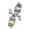
| ||||||||
|---|---|---|---|---|---|---|---|---|---|
| 1 | 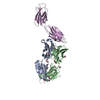
| ||||||||
| 2 | 
| ||||||||
| Unit cell |
|
-Components
| #1: Antibody | Mass: 24388.352 Da / Num. of mol.: 2 Source method: isolated from a genetically manipulated source Source: (gene. exp.) Homo sapiens (human) / Production host:  #2: Antibody | Mass: 23415.867 Da / Num. of mol.: 2 Source method: isolated from a genetically manipulated source Source: (gene. exp.) Homo sapiens (human) / Production host: #3: Protein | Mass: 22355.982 Da / Num. of mol.: 2 Source method: isolated from a genetically manipulated source Source: (gene. exp.) Homo sapiens (human) / Gene: TEK, TIE2, VMCM, VMCM1 / Production host: References: UniProt: Q02763, receptor protein-tyrosine kinase #4: Chemical | ChemComp-EDO /   Mass: 62.068 Da / Num. of mol.: 34 / Source method: obtained synthetically / Formula: C2H6O2 Mass: 62.068 Da / Num. of mol.: 34 / Source method: obtained synthetically / Formula: C2H6O2#5: Water | ChemComp-HOH / |  Mass: 18.015 Da / Num. of mol.: 737 / Source method: isolated from a natural source / Formula: H2O Mass: 18.015 Da / Num. of mol.: 737 / Source method: isolated from a natural source / Formula: H2OHas ligand of interest | N | Has protein modification | Y | |
|---|
-Experimental details
-Experiment
| Experiment | Method: X-RAY DIFFRACTION / Number of used crystals: 1 |
|---|
- Sample preparation
| Crystal | Density Matthews: 2.92 Å3/Da / Density % sol: 57.82 % |
|---|---|
| Crystal grow | Temperature: 292.15 K / Method: vapor diffusion, hanging drop / pH: 8.5 Details: 100 mM Bis-Tris propane pH 8.5, 110 mM potassium dihydrogen phosphate, 15% (w/v) polyethylene glycol 3350 PH range: 8.5 |
-Data collection
| Diffraction | Mean temperature: 100 K / Serial crystal experiment: N |
|---|---|
| Diffraction source | Source: SYNCHROTRON / Site: PAL/PLS / Beamline: 11C / Wavelength: 0.9794 Å |
| Detector | Type: DECTRIS PILATUS3 S 6M / Detector: PIXEL / Date: Jun 25, 2019 |
| Radiation | Protocol: SINGLE WAVELENGTH / Monochromatic (M) / Laue (L): M / Scattering type: x-ray |
| Radiation wavelength | Wavelength: 0.9794 Å / Relative weight: 1 |
| Reflection | Resolution: 2.094→44.72 Å / Num. obs: 84953 / % possible obs: 91.04 % / Redundancy: 3.6 % / Biso Wilson estimate: 25.96 Å2 / Rmerge(I) obs: 0.099 / Net I/σ(I): 7.126 |
| Reflection shell | Resolution: 2.094→2.169 Å / Redundancy: 3.4 % / Rmerge(I) obs: 0.509 / Mean I/σ(I) obs: 1.217 / Num. unique obs: 8247 / % possible all: 88.61 |
- Processing
| Software |
| ||||||||||||||||||||||||||||||||||||||||||||||||||||||||||||||||||||||||||||||||||||
|---|---|---|---|---|---|---|---|---|---|---|---|---|---|---|---|---|---|---|---|---|---|---|---|---|---|---|---|---|---|---|---|---|---|---|---|---|---|---|---|---|---|---|---|---|---|---|---|---|---|---|---|---|---|---|---|---|---|---|---|---|---|---|---|---|---|---|---|---|---|---|---|---|---|---|---|---|---|---|---|---|---|---|---|---|---|
| Refinement | Method to determine structure: MOLECULAR REPLACEMENT Starting model: 5X8M, 5MYA Resolution: 2.094→44.72 Å / SU ML: 0.25 / Cross valid method: THROUGHOUT / σ(F): 1.96 / Phase error: 23.52 / Stereochemistry target values: ML
| ||||||||||||||||||||||||||||||||||||||||||||||||||||||||||||||||||||||||||||||||||||
| Solvent computation | Shrinkage radii: 0.9 Å / VDW probe radii: 1.11 Å / Solvent model: FLAT BULK SOLVENT MODEL | ||||||||||||||||||||||||||||||||||||||||||||||||||||||||||||||||||||||||||||||||||||
| Displacement parameters | Biso max: 132.41 Å2 / Biso mean: 39.2863 Å2 / Biso min: 11.53 Å2 | ||||||||||||||||||||||||||||||||||||||||||||||||||||||||||||||||||||||||||||||||||||
| Refinement step | Cycle: final / Resolution: 2.094→44.72 Å
| ||||||||||||||||||||||||||||||||||||||||||||||||||||||||||||||||||||||||||||||||||||
| LS refinement shell | Refine-ID: X-RAY DIFFRACTION / Rfactor Rfree error: 0
| ||||||||||||||||||||||||||||||||||||||||||||||||||||||||||||||||||||||||||||||||||||
| Refinement TLS params. | Method: refined / Origin x: 34.9749 Å / Origin y: 15.1799 Å / Origin z: 89.5922 Å
| ||||||||||||||||||||||||||||||||||||||||||||||||||||||||||||||||||||||||||||||||||||
| Refinement TLS group |
|
