 Movie
Movie Controller

[English] 日本語
 Yorodumi
- PDB-7e6t: Structural insights into the activation of human calcium-sensing ... -
+ Open data

- Basic information
| Entry | Database: PDB / ID: 7e6t | |||||||||||||||||||||
|---|---|---|---|---|---|---|---|---|---|---|---|---|---|---|---|---|---|---|---|---|---|---|
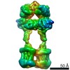
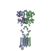
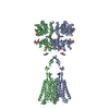
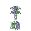
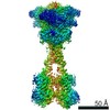
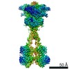
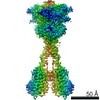
| Title | Structural insights into the activation of human calcium-sensing receptor | |||||||||||||||||||||
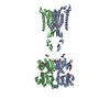
 Components Components | Extracellular calcium-sensing receptor | |||||||||||||||||||||
 Keywords Keywords | STRUCTURAL PROTEIN / G-protein-coupled receptor (GPCR) / Calcium-sensing receptor (CaSR) / cryo-electron microscopy (cryo-EM) / calcium ions / nanobody | |||||||||||||||||||||
| Function / homology |  Function and homology information Function and homology informationregulation of presynaptic membrane potential / bile acid secretion / response to fibroblast growth factor / chemosensory behavior / cellular response to peptide / cellular response to vitamin D / Class C/3 (Metabotropic glutamate/pheromone receptors) / positive regulation of positive chemotaxis / cellular response to hepatocyte growth factor stimulus / fat pad development ...regulation of presynaptic membrane potential / bile acid secretion / response to fibroblast growth factor / chemosensory behavior / cellular response to peptide / cellular response to vitamin D / Class C/3 (Metabotropic glutamate/pheromone receptors) / positive regulation of positive chemotaxis / cellular response to hepatocyte growth factor stimulus / fat pad development / branching morphogenesis of an epithelial tube / amino acid binding / positive regulation of calcium ion import / regulation of calcium ion transport / positive regulation of NLRP3 inflammasome complex assembly / cellular response to low-density lipoprotein particle stimulus / positive regulation of vasoconstriction / anatomical structure morphogenesis / detection of calcium ion / JNK cascade / response to ischemia / ossification / axon terminus / chloride transmembrane transport / cellular response to glucose stimulus / integrin binding / vasodilation / intracellular calcium ion homeostasis / G protein-coupled receptor activity / positive regulation of insulin secretion / adenylate cyclase-inhibiting G protein-coupled receptor signaling pathway / presynaptic membrane / cellular response to hypoxia / G alpha (i) signalling events / basolateral plasma membrane / G alpha (q) signalling events / phospholipase C-activating G protein-coupled receptor signaling pathway / transmembrane transporter binding / positive regulation of ERK1 and ERK2 cascade / apical plasma membrane / G protein-coupled receptor signaling pathway / neuronal cell body / calcium ion binding / positive regulation of cell population proliferation / positive regulation of gene expression / protein kinase binding / glutamatergic synapse / cell surface / protein homodimerization activity / identical protein binding / plasma membrane Similarity search - Function | |||||||||||||||||||||
| Biological species |  Homo sapiens (human) Homo sapiens (human) | |||||||||||||||||||||
| Method | ELECTRON MICROSCOPY / single particle reconstruction / cryo EM / Resolution: 3 Å | |||||||||||||||||||||
 Authors Authors | Geng, Y. / Chen, X.C. / Wang, L. / Cui, Q.Q. / Ding, Z.Y. / Han, L. / Kou, Y.J. / Zhang, W.Q. / Wang, H.N. / Jia, X.M. ...Geng, Y. / Chen, X.C. / Wang, L. / Cui, Q.Q. / Ding, Z.Y. / Han, L. / Kou, Y.J. / Zhang, W.Q. / Wang, H.N. / Jia, X.M. / Dai, M. / Shi, Z.Z. / Li, Y.Y. / Li, X.Y. | |||||||||||||||||||||
| Funding support |  China, 2items China, 2items
| |||||||||||||||||||||
 Citation Citation | Journal: Elife / Year: 2021 Title: Structural insights into the activation of human calcium-sensing receptor. Authors: Xiaochen Chen / Lu Wang / Qianqian Cui / Zhanyu Ding / Li Han / Yongjun Kou / Wenqing Zhang / Haonan Wang / Xiaomin Jia / Mei Dai / Zhenzhong Shi / Yuying Li / Xiyang Li / Yong Geng / Abstract: Human calcium-sensing receptor (CaSR) is a G-protein-coupled receptor that maintains Ca homeostasis in serum. Here, we present the cryo-electron microscopy structures of the CaSR in the inactive and ...Human calcium-sensing receptor (CaSR) is a G-protein-coupled receptor that maintains Ca homeostasis in serum. Here, we present the cryo-electron microscopy structures of the CaSR in the inactive and agonist+PAM bound states. Complemented with previously reported structures of CaSR, we show that in addition to the full inactive and active states, there are multiple intermediate states during the activation of CaSR. We used a negative allosteric nanobody to stabilize the CaSR in the fully inactive state and found a new binding site for Ca ion that acts as a composite agonist with L-amino acid to stabilize the closure of active Venus flytraps. Our data show that agonist binding leads to compaction of the dimer, proximity of the cysteine-rich domains, large-scale transitions of seven-transmembrane domains, and inter- and intrasubunit conformational changes of seven-transmembrane domains to accommodate downstream transducers. Our results reveal the structural basis for activation mechanisms of CaSR and clarify the mode of action of Ca ions and L-amino acid leading to the activation of the receptor. | |||||||||||||||||||||
| History |
|
- Structure visualization
| Movie |
Movie viewer |
|---|---|
| Structure viewer | Molecule: MolmilJmol/JSmol |
- Downloads & links
-Download
| PDBx/mmCIF format | 7e6t.cif.gz | 294.3 KB | Display | PDBx/mmCIF format |
|---|---|---|---|---|
| PDB format | pdb7e6t.ent.gz | 232.4 KB | Display | PDB format |
| PDBx/mmJSON format | 7e6t.json.gz | Tree view | PDBx/mmJSON format | |
| Others |  Other downloads Other downloads |
-Validation report
| Arichive directory | https://data.pdbj.org/pub/pdb/validation_reports/e6/7e6tftp://data.pdbj.org/pub/pdb/validation_reports/e6/7e6t | HTTPS FTP |
|---|
-Related structure data
| Related structure data | 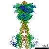 30996MC  7e6uC M: map data used to model this data C: citing same article ( |
|---|---|
| Similar structure data |

-Links
 PDBj
PDBj


- Assembly
| Deposited unit | 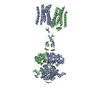
| |||||||||||||||||||||||||||
|---|---|---|---|---|---|---|---|---|---|---|---|---|---|---|---|---|---|---|---|---|---|---|---|---|---|---|---|---|
| 1 |
| |||||||||||||||||||||||||||
| Noncrystallographic symmetry (NCS) | NCS domain:
NCS domain segments:
|
-Components
| #1: Protein | Mass: 97326.094 Da / Num. of mol.: 2 Source method: isolated from a genetically manipulated source Source: (gene. exp.) Homo sapiens (human) / Gene: CASR, GPRC2A, PCAR1 / Production host: Homo sapiens (human) / References: UniProt: P41180#2: Chemical |   Mass: 94.971 Da / Num. of mol.: 2 / Source method: obtained synthetically / Formula: PO4 / Feature type: SUBJECT OF INVESTIGATION Mass: 94.971 Da / Num. of mol.: 2 / Source method: obtained synthetically / Formula: PO4 / Feature type: SUBJECT OF INVESTIGATION#3: Chemical | ChemComp-CA /   Mass: 40.078 Da / Num. of mol.: 6 / Source method: obtained synthetically / Formula: Ca / Feature type: SUBJECT OF INVESTIGATION Mass: 40.078 Da / Num. of mol.: 6 / Source method: obtained synthetically / Formula: Ca / Feature type: SUBJECT OF INVESTIGATION#4: Sugar | ChemComp-NAG /   Type: D-saccharide, beta linking / Mass: 221.208 Da / Num. of mol.: 4 / Source method: obtained synthetically / Formula: C8H15NO6 / Feature type: SUBJECT OF INVESTIGATION Type: D-saccharide, beta linking / Mass: 221.208 Da / Num. of mol.: 4 / Source method: obtained synthetically / Formula: C8H15NO6 / Feature type: SUBJECT OF INVESTIGATION#5: Chemical | 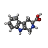  Type: L-peptide linking / Mass: 216.236 Da / Num. of mol.: 2 / Source method: obtained synthetically / Formula: C12H12N2O2 / Feature type: SUBJECT OF INVESTIGATION Type: L-peptide linking / Mass: 216.236 Da / Num. of mol.: 2 / Source method: obtained synthetically / Formula: C12H12N2O2 / Feature type: SUBJECT OF INVESTIGATIONHas ligand of interest | Y | Has protein modification | Y | |
|---|
-Experimental details
-Experiment
| Experiment | Method: ELECTRON MICROSCOPY |
|---|---|
| EM experiment | Aggregation state: 2D ARRAY / 3D reconstruction method: single particle reconstruction |
- Sample preparation
| Component | Name: CaSR, TNCA, Ca, PO4 / Type: COMPLEX / Entity ID: #1 / Source: RECOMBINANT |
|---|---|
| Source (natural) | Organism: Homo sapiens (human) |
| Source (recombinant) | Organism: Homo sapiens (human) |
| Buffer solution | pH: 8 |
| Specimen | Embedding applied: NO / Shadowing applied: NO / Staining applied: NO / Vitrification applied: YES |
| Vitrification | Cryogen name: NITROGEN |
- Electron microscopy imaging
| Experimental equipment |  Model: Titan Krios / Image courtesy: FEI Company |
|---|---|
| Microscopy | Model: FEI TITAN KRIOS |
| Electron gun | Electron source:  FIELD EMISSION GUN / Accelerating voltage: 300 kV / Illumination mode: OTHER FIELD EMISSION GUN / Accelerating voltage: 300 kV / Illumination mode: OTHER |
| Electron lens | Mode: BRIGHT FIELD / Alignment procedure: ZEMLIN TABLEAU |
| Specimen holder | Cryogen: NITROGEN / Specimen holder model: SIDE ENTRY, EUCENTRIC |
| Image recording | Electron dose: 70 e/Å2 / Film or detector model: GATAN K3 BIOQUANTUM (6k x 4k) |
| EM imaging optics | Phase plate: ZERNIKE PHASE PLATE |
- Processing
| Software | Name: PHENIX / Version: 1.18.2_3874: / Classification: refinement | ||||||||||||||||||||||||
|---|---|---|---|---|---|---|---|---|---|---|---|---|---|---|---|---|---|---|---|---|---|---|---|---|---|
| EM software | Name: PHENIX / Category: model refinement | ||||||||||||||||||||||||
| CTF correction | Type: PHASE FLIPPING AND AMPLITUDE CORRECTION | ||||||||||||||||||||||||
| 3D reconstruction | Resolution: 3 Å / Resolution method: FSC 0.143 CUT-OFF / Num. of particles: 248900 / Symmetry type: POINT | ||||||||||||||||||||||||
| Refinement | Resolution: 3→353.6 Å / Cor.coef. Fo:Fc: 0.672 / SU B: 8.559 / SU ML: 0.117 / ESU R: 0.093 Stereochemistry target values: MAXIMUM LIKELIHOOD WITH PHASES
| ||||||||||||||||||||||||
| Solvent computation | Solvent model: PARAMETERS FOR MASK CACLULATION | ||||||||||||||||||||||||
| Displacement parameters | Biso mean: 179.014 Å2
| ||||||||||||||||||||||||
| Refinement step | Cycle: 1 / Total: 12766 | ||||||||||||||||||||||||
| Refine LS restraints |
| ||||||||||||||||||||||||
| Refine LS restraints NCS | Ens-ID: 1 / Number: 43116 / Refine-ID: ELECTRON MICROSCOPY / Type: interatomic distance / Rms dev position: 0.21 Å / Weight position: 0.05
| ||||||||||||||||||||||||
| LS refinement shell | Resolution: 3→3.078 Å / Total num. of bins used: 20
|
