 Movie
Movie Controller

[English] 日本語
 Yorodumi
- PDB-7e5g: HUMAN PPAR ALPHA LIGAND BINDING DOMAIN IN COMPLEX WITH YN4pai OBT... -
+ Open data

- Basic information
| Entry | Database: PDB / ID: 7e5g | ||||||
|---|---|---|---|---|---|---|---|
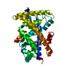
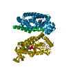
| Title | HUMAN PPAR ALPHA LIGAND BINDING DOMAIN IN COMPLEX WITH YN4pai OBTAINED BY SOAKING | ||||||
 Components Components | Peroxisome proliferator-activated receptor alpha | ||||||
 Keywords Keywords | TRANSCRIPTION / NUCLEAR RECEPTOR / PPAR / PROTEIN-LIGAND COMPLEX | ||||||
| Function / homology |  Function and homology information Function and homology informationregulation of fatty acid transport / enamel mineralization / positive regulation of fatty acid beta-oxidation / regulation of ketone metabolic process / cellular response to fructose stimulus / negative regulation of hepatocyte apoptotic process / negative regulation of cell growth involved in cardiac muscle cell development / regulation of fatty acid metabolic process / positive regulation of fatty acid oxidation / behavioral response to nicotine ...regulation of fatty acid transport / enamel mineralization / positive regulation of fatty acid beta-oxidation / regulation of ketone metabolic process / cellular response to fructose stimulus / negative regulation of hepatocyte apoptotic process / negative regulation of cell growth involved in cardiac muscle cell development / regulation of fatty acid metabolic process / positive regulation of fatty acid oxidation / behavioral response to nicotine / negative regulation of appetite / lipoprotein metabolic process / negative regulation of leukocyte cell-cell adhesion / mitogen-activated protein kinase kinase kinase binding / ubiquitin conjugating enzyme binding / negative regulation of glycolytic process / positive regulation of fatty acid metabolic process / NFAT protein binding / DNA-binding transcription activator activity / nuclear steroid receptor activity / negative regulation of cholesterol storage / epidermis development / positive regulation of ATP biosynthetic process / negative regulation of macrophage derived foam cell differentiation / positive regulation of lipid biosynthetic process / phosphatase binding / Transcriptional regulation of brown and beige adipocyte differentiation by EBF2 / intracellular receptor signaling pathway / nitric oxide metabolic process / negative regulation of reactive oxygen species biosynthetic process / peroxisome proliferator activated receptor signaling pathway / hormone-mediated signaling pathway / Regulation of lipid metabolism by PPARalpha / negative regulation of blood pressure / lactation / BMAL1:CLOCK,NPAS2 activates circadian expression / negative regulation of cytokine production involved in inflammatory response / response to nutrient / RORA,B,C and NR1D1 (REV-ERBA) regulate gene expression / Activation of gene expression by SREBF (SREBP) / Expression of BMAL (ARNTL), CLOCK, and NPAS2 / MDM2/MDM4 family protein binding / negative regulation of miRNA transcription / positive regulation of gluconeogenesis / fatty acid metabolic process / negative regulation of phosphatidylinositol 3-kinase/protein kinase B signal transduction / cellular response to starvation / gluconeogenesis / SUMOylation of intracellular receptors / circadian regulation of gene expression / wound healing / Heme signaling / negative regulation of transforming growth factor beta receptor signaling pathway / PPARA activates gene expression / Transcriptional activation of mitochondrial biogenesis / Cytoprotection by HMOX1 / Nuclear Receptor transcription pathway / Transcriptional regulation of white adipocyte differentiation / response to insulin / regulation of circadian rhythm / negative regulation of inflammatory response / DNA-binding transcription repressor activity, RNA polymerase II-specific / nuclear receptor activity / RNA polymerase II transcription regulator complex / transcription coactivator binding / heart development / gene expression / DNA-binding transcription activator activity, RNA polymerase II-specific / sequence-specific DNA binding / defense response to virus / RNA polymerase II-specific DNA-binding transcription factor binding / response to ethanol / DNA-binding transcription factor activity, RNA polymerase II-specific / response to hypoxia / cell differentiation / RNA polymerase II cis-regulatory region sequence-specific DNA binding / protein ubiquitination / DNA-binding transcription factor activity / protein domain specific binding / lipid binding / positive regulation of DNA-templated transcription / chromatin / protein-containing complex binding / negative regulation of transcription by RNA polymerase II / positive regulation of transcription by RNA polymerase II / DNA binding / zinc ion binding / nucleoplasm / nucleus Similarity search - Function | ||||||
| Biological species |  Homo sapiens (human) Homo sapiens (human) | ||||||
| Method |  X-RAY DIFFRACTION / SYNCHROTRON / MOLECULAR REPLACEMENT / Resolution: 1.66 Å X-RAY DIFFRACTION / SYNCHROTRON / MOLECULAR REPLACEMENT / Resolution: 1.66 Å | ||||||
 Authors Authors | Oyama, T. / Kamata, S. / Ishii, I. / Miyachi, H. | ||||||
 Citation Citation | Journal: Biol.Pharm.Bull. / Year: 2021 Title: Crystal Structures of the Human Peroxisome Proliferator-Activated Receptor (PPAR) alpha Ligand-Binding Domain in Complexes with a Series of Phenylpropanoic Acid Derivatives Generated by a ...Title: Crystal Structures of the Human Peroxisome Proliferator-Activated Receptor (PPAR) alpha Ligand-Binding Domain in Complexes with a Series of Phenylpropanoic Acid Derivatives Generated by a Ligand-Exchange Soaking Method. Authors: Oyama, T. / Kamata, S. / Ishii, I. / Miyachi, H. | ||||||
| History |
|
- Structure visualization
| Structure viewer | Molecule: MolmilJmol/JSmol |
|---|
- Downloads & links
-Download
| PDBx/mmCIF format | 7e5g.cif.gz | 85.2 KB | Display | PDBx/mmCIF format |
|---|---|---|---|---|
| PDB format | pdb7e5g.ent.gz | 50.2 KB | Display | PDB format |
| PDBx/mmJSON format | 7e5g.json.gz | Tree view | PDBx/mmJSON format | |
| Others |  Other downloads Other downloads |
-Validation report
| Arichive directory | https://data.pdbj.org/pub/pdb/validation_reports/e5/7e5gftp://data.pdbj.org/pub/pdb/validation_reports/e5/7e5g | HTTPS FTP |
|---|
-Related structure data
| Related structure data |  7e5fC 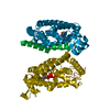 7e5hC  7e5iC 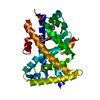 2znnS S: Starting model for refinement C: citing same article ( |
|---|---|
| Similar structure data |


-Links
 PDBj
PDBj


- Assembly
| Deposited unit | 
| ||||||||||||
|---|---|---|---|---|---|---|---|---|---|---|---|---|---|
| 1 | 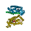
| ||||||||||||
| Unit cell |
|
-Components
| #1: Protein | Mass: 30856.053 Da / Num. of mol.: 1 Source method: isolated from a genetically manipulated source Source: (gene. exp.) Homo sapiens (human) / Production host:  |
|---|---|
| #2: Chemical | ChemComp-HVX / (  Mass: 507.619 Da / Num. of mol.: 1 / Source method: obtained synthetically / Formula: C33H33NO4 / Feature type: SUBJECT OF INVESTIGATION Mass: 507.619 Da / Num. of mol.: 1 / Source method: obtained synthetically / Formula: C33H33NO4 / Feature type: SUBJECT OF INVESTIGATION |
| #3: Water | ChemComp-HOH /  Mass: 18.015 Da / Num. of mol.: 113 / Source method: isolated from a natural source / Formula: H2O Mass: 18.015 Da / Num. of mol.: 113 / Source method: isolated from a natural source / Formula: H2O |
| Has ligand of interest | Y |
-Experimental details
-Experiment
| Experiment | Method: X-RAY DIFFRACTION / Number of used crystals: 1 |
|---|
- Sample preparation
| Crystal | Density Matthews: 2.29 Å3/Da / Density % sol: 46.33 % |
|---|---|
| Crystal grow | Temperature: 277 K / Method: vapor diffusion, hanging drop / pH: 6.5 Details: 0.1 M Bis-Tris, 25% (w/v) polyethylene glycol (PEG) 3350 |
-Data collection
| Diffraction | Mean temperature: 100 K / Serial crystal experiment: N |
|---|---|
| Diffraction source | Source: SYNCHROTRON / Site: SPring-8  / Beamline: BL26B1 / Wavelength: 1 Å / Beamline: BL26B1 / Wavelength: 1 Å |
| Detector | Type: DECTRIS EIGER X 4M / Detector: PIXEL / Date: Apr 12, 2017 / Details: two dimensional focusing mirror |
| Radiation | Monochromator: fixed exit Si double crystal monochromator / Protocol: SINGLE WAVELENGTH / Monochromatic (M) / Laue (L): M / Scattering type: x-ray |
| Radiation wavelength | Wavelength: 1 Å / Relative weight: 1 |
| Reflection | Resolution: 1.66→50 Å / Num. obs: 63155 / % possible obs: 99.9 % / Redundancy: 3.4 % / Biso Wilson estimate: 17.06 Å2 / CC1/2: 0.997 / Rmerge(I) obs: 0.046 / Rpim(I) all: 0.044 / Rrim(I) all: 0.063 / Net I/σ(I): 14.1 |
| Reflection shell | Resolution: 1.66→1.69 Å / Redundancy: 3.5 % / Rmerge(I) obs: 0.321 / Mean I/σ(I) obs: 3.8 / Num. unique obs: 1590 / CC1/2: 0.906 / Rpim(I) all: 0.3 / Rrim(I) all: 0.44 / % possible all: 100 |
- Processing
| Software |
| |||||||||||||||||||||||||||||||||||||||||||||||||||||||||||||||||||||||||||||||||||||||||||||||||||||||||||||||||||||||||||||||||||||||||||||||||||||||||||||||||
|---|---|---|---|---|---|---|---|---|---|---|---|---|---|---|---|---|---|---|---|---|---|---|---|---|---|---|---|---|---|---|---|---|---|---|---|---|---|---|---|---|---|---|---|---|---|---|---|---|---|---|---|---|---|---|---|---|---|---|---|---|---|---|---|---|---|---|---|---|---|---|---|---|---|---|---|---|---|---|---|---|---|---|---|---|---|---|---|---|---|---|---|---|---|---|---|---|---|---|---|---|---|---|---|---|---|---|---|---|---|---|---|---|---|---|---|---|---|---|---|---|---|---|---|---|---|---|---|---|---|---|---|---|---|---|---|---|---|---|---|---|---|---|---|---|---|---|---|---|---|---|---|---|---|---|---|---|---|---|---|---|---|---|
| Refinement | Method to determine structure: MOLECULAR REPLACEMENT Starting model: 2znn Resolution: 1.66→43.07 Å / SU ML: 0.207 / Cross valid method: FREE R-VALUE / σ(F): 0.02 / Phase error: 24.9067 Stereochemistry target values: GeoStd + Monomer Library + CDL v1.2
| |||||||||||||||||||||||||||||||||||||||||||||||||||||||||||||||||||||||||||||||||||||||||||||||||||||||||||||||||||||||||||||||||||||||||||||||||||||||||||||||||
| Solvent computation | Shrinkage radii: 0.9 Å / VDW probe radii: 1.11 Å / Solvent model: FLAT BULK SOLVENT MODEL | |||||||||||||||||||||||||||||||||||||||||||||||||||||||||||||||||||||||||||||||||||||||||||||||||||||||||||||||||||||||||||||||||||||||||||||||||||||||||||||||||
| Displacement parameters | Biso mean: 20.79 Å2 | |||||||||||||||||||||||||||||||||||||||||||||||||||||||||||||||||||||||||||||||||||||||||||||||||||||||||||||||||||||||||||||||||||||||||||||||||||||||||||||||||
| Refinement step | Cycle: LAST / Resolution: 1.66→43.07 Å
| |||||||||||||||||||||||||||||||||||||||||||||||||||||||||||||||||||||||||||||||||||||||||||||||||||||||||||||||||||||||||||||||||||||||||||||||||||||||||||||||||
| Refine LS restraints |
| |||||||||||||||||||||||||||||||||||||||||||||||||||||||||||||||||||||||||||||||||||||||||||||||||||||||||||||||||||||||||||||||||||||||||||||||||||||||||||||||||
| LS refinement shell |
|
