 Movie
Movie Controller

[English] 日本語
 Yorodumi
- PDB-2znn: Human PPAR alpha ligand binding domain in complex with a syntheti... -
+ Open data

- Basic information
| Entry | Database: PDB / ID: 2znn | ||||||
|---|---|---|---|---|---|---|---|
| Title | Human PPAR alpha ligand binding domain in complex with a synthetic agonist TIPP703 | ||||||
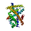
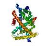
 Components Components | Peroxisome proliferator-activated receptor alpha | ||||||
 Keywords Keywords | TRANSCRIPTION / NUCLEAR RECEPTOR / PROTEIN-LIGAND COMPLEX / PPAR / Activator / DNA-binding / Metal-binding / Nucleus / Receptor / Transcription regulation / Zinc-finger | ||||||
| Function / homology |  Function and homology information Function and homology informationregulation of fatty acid transport / enamel mineralization / positive regulation of fatty acid beta-oxidation / regulation of ketone metabolic process / cellular response to fructose stimulus / negative regulation of cell growth involved in cardiac muscle cell development / regulation of fatty acid metabolic process / negative regulation of appetite / negative regulation of hepatocyte apoptotic process / positive regulation of fatty acid oxidation ...regulation of fatty acid transport / enamel mineralization / positive regulation of fatty acid beta-oxidation / regulation of ketone metabolic process / cellular response to fructose stimulus / negative regulation of cell growth involved in cardiac muscle cell development / regulation of fatty acid metabolic process / negative regulation of appetite / negative regulation of hepatocyte apoptotic process / positive regulation of fatty acid oxidation / behavioral response to nicotine / lipoprotein metabolic process / negative regulation of leukocyte cell-cell adhesion / mitogen-activated protein kinase kinase kinase binding / ubiquitin conjugating enzyme binding / negative regulation of glycolytic process / positive regulation of fatty acid metabolic process / DNA-binding transcription activator activity / NFAT protein binding / negative regulation of cholesterol storage / nuclear steroid receptor activity / positive regulation of ATP biosynthetic process / negative regulation of macrophage derived foam cell differentiation / epidermis development / positive regulation of lipid biosynthetic process / phosphatase binding / Transcriptional regulation of brown and beige adipocyte differentiation by EBF2 / intracellular receptor signaling pathway / nitric oxide metabolic process / negative regulation of reactive oxygen species biosynthetic process / Regulation of lipid metabolism by PPARalpha / negative regulation of blood pressure / lactation / peroxisome proliferator activated receptor signaling pathway / hormone-mediated signaling pathway / BMAL1:CLOCK,NPAS2 activates circadian expression / response to nutrient / negative regulation of cytokine production involved in inflammatory response / RORA,B,C and NR1D1 (REV-ERBA) regulate gene expression / Activation of gene expression by SREBF (SREBP) / Expression of BMAL (ARNTL), CLOCK, and NPAS2 / MDM2/MDM4 family protein binding / negative regulation of miRNA transcription / positive regulation of gluconeogenesis / negative regulation of phosphatidylinositol 3-kinase/protein kinase B signal transduction / cellular response to starvation / gluconeogenesis / SUMOylation of intracellular receptors / circadian regulation of gene expression / wound healing / Heme signaling / negative regulation of transforming growth factor beta receptor signaling pathway / PPARA activates gene expression / Transcriptional activation of mitochondrial biogenesis / Cytoprotection by HMOX1 / fatty acid metabolic process / Nuclear Receptor transcription pathway / Transcriptional regulation of white adipocyte differentiation / regulation of circadian rhythm / response to insulin / DNA-binding transcription repressor activity, RNA polymerase II-specific / nuclear receptor activity / negative regulation of inflammatory response / RNA polymerase II transcription regulator complex / transcription coactivator binding / heart development / DNA-binding transcription activator activity, RNA polymerase II-specific / sequence-specific DNA binding / gene expression / defense response to virus / RNA polymerase II-specific DNA-binding transcription factor binding / response to ethanol / DNA-binding transcription factor activity, RNA polymerase II-specific / response to hypoxia / cell differentiation / RNA polymerase II cis-regulatory region sequence-specific DNA binding / protein ubiquitination / DNA-binding transcription factor activity / protein domain specific binding / lipid binding / positive regulation of DNA-templated transcription / chromatin / protein-containing complex binding / negative regulation of transcription by RNA polymerase II / positive regulation of transcription by RNA polymerase II / DNA binding / zinc ion binding / nucleoplasm / nucleus Similarity search - Function | ||||||
| Biological species |  Homo sapiens (human) Homo sapiens (human) | ||||||
| Method |  X-RAY DIFFRACTION / SYNCHROTRON / MOLECULAR REPLACEMENT / Resolution: 2.01 Å X-RAY DIFFRACTION / SYNCHROTRON / MOLECULAR REPLACEMENT / Resolution: 2.01 Å | ||||||
 Authors Authors | Oyama, T. / Toyota, K. / Kasuga, J. / Miyachi, H. / Morikawa, K. | ||||||
 Citation Citation | Journal: Acta Crystallogr.,Sect.D / Year: 2009 Title: Adaptability and selectivity of human peroxisome proliferator-activated receptor (PPAR) pan agonists revealed from crystal structures Authors: Oyama, T. / Toyota, K. / Waku, T. / Hirakawa, Y. / Nagasawa, N. / Kasuga, J. / Hashimoto, Y. / Miyachi, H. / Morikawa, K. #1: Journal: To be PublishedTitle: Peroxisome proliferator-activated receptor (PPAR) agonists with a 3,4-dihydro-2H-benzo[e][1,3]oxazine and 2,3,4,5-tetrahydrobenzo[f][1,4]oxazepine skeleton: effects of the conformational ...Title: Peroxisome proliferator-activated receptor (PPAR) agonists with a 3,4-dihydro-2H-benzo[e][1,3]oxazine and 2,3,4,5-tetrahydrobenzo[f][1,4]oxazepine skeleton: effects of the conformational restriction on PPAR agonistic acivity Authors: Ohkane, K. / Kasuga, J. / Oyama, T. / Hirakawa, Y. / Morikawa, K. / Makishima, M. / Hashimoto, Y. / Miyachi, H. #2: Journal: To be PublishedTitle: Improvement of the transactivation activity of phenylpropanoic acid-type peroxisome proliferator-acitvated rectpror pan agonists; effect of introduction of fluorine at the linker part Authors: Kasuga, J. / Oyama, T. / Hirakawa, Y. / Makishima, M. / Morikawa, K. / Hashimoto, Y. / Miyachi, H. | ||||||
| History |
|
- Structure visualization
| Structure viewer | Molecule: MolmilJmol/JSmol |
|---|
- Downloads & links
-Download
| PDBx/mmCIF format | 2znn.cif.gz | 69.9 KB | Display | PDBx/mmCIF format |
|---|---|---|---|---|
| PDB format | pdb2znn.ent.gz | 49.6 KB | Display | PDB format |
| PDBx/mmJSON format | 2znn.json.gz | Tree view | PDBx/mmJSON format | |
| Others |  Other downloads Other downloads |
-Validation report
| Arichive directory | https://data.pdbj.org/pub/pdb/validation_reports/zn/2znnftp://data.pdbj.org/pub/pdb/validation_reports/zn/2znn | HTTPS FTP |
|---|
-Related structure data
| Related structure data | 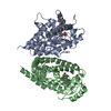 2znoC  2znpC  2znqC 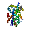 1i7gS C: citing same article ( S: Starting model for refinement |
|---|---|
| Similar structure data |


-Links
 PDBj
PDBj


- Assembly
| Deposited unit | 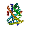
| ||||||||
|---|---|---|---|---|---|---|---|---|---|
| 1 |
| ||||||||
| Unit cell |
|
-Components
| #1: Protein | Mass: 30856.053 Da / Num. of mol.: 1 / Fragment: Ligand binding domain Source method: isolated from a genetically manipulated source Source: (gene. exp.) Homo sapiens (human) / Gene: PPARA, NR1C1, PPAR / Plasmid: pET28a / Production host:  |
|---|---|
| #2: Chemical | ChemComp-S44 / (  Mass: 503.672 Da / Num. of mol.: 1 / Source method: obtained synthetically / Formula: C32H41NO4 Mass: 503.672 Da / Num. of mol.: 1 / Source method: obtained synthetically / Formula: C32H41NO4 |
| #3: Water | ChemComp-HOH /  Mass: 18.015 Da / Num. of mol.: 146 / Source method: isolated from a natural source / Formula: H2O Mass: 18.015 Da / Num. of mol.: 146 / Source method: isolated from a natural source / Formula: H2O |
-Experimental details
-Experiment
| Experiment | Method: X-RAY DIFFRACTION / Number of used crystals: 1 |
|---|
- Sample preparation
| Crystal | Density Matthews: 2.26 Å3/Da / Density % sol: 45.47 % |
|---|---|
| Crystal grow | Temperature: 293 K / Method: vapor diffusion, hanging drop / pH: 7.5 Details: 25% PEG3350, 0.1M HEPES, pH 7.5, VAPOR DIFFUSION, HANGING DROP, temperature 293K |
-Data collection
| Diffraction | Mean temperature: 100 K |
|---|---|
| Diffraction source | Source: SYNCHROTRON / Site: SPring-8  / Beamline: BL38B1 / Wavelength: 1 Å / Beamline: BL38B1 / Wavelength: 1 Å |
| Detector | Type: RIGAKU JUPITER 210 / Detector: CCD / Date: Mar 13, 2007 |
| Radiation | Monochromator: Fixed exit Si 111 double crystal monochromator Protocol: SINGLE WAVELENGTH / Monochromatic (M) / Laue (L): M / Scattering type: x-ray |
| Radiation wavelength | Wavelength: 1 Å / Relative weight: 1 |
| Reflection | Resolution: 2→50 Å / Num. obs: 18142 / % possible obs: 98.7 % / Observed criterion σ(F): 0 / Redundancy: 3.7 % / Biso Wilson estimate: 11.6 Å2 / Rmerge(I) obs: 0.064 / Net I/σ(I): 10.6 |
| Reflection shell | Resolution: 2→2.07 Å / Redundancy: 3.1 % / Rmerge(I) obs: 0.222 / Num. unique all: 1646 / % possible all: 90.8 |
- Processing
| Software |
| ||||||||||||||||||||||||||||||||||||
|---|---|---|---|---|---|---|---|---|---|---|---|---|---|---|---|---|---|---|---|---|---|---|---|---|---|---|---|---|---|---|---|---|---|---|---|---|---|
| Refinement | Method to determine structure: MOLECULAR REPLACEMENT Starting model: pdb entry 1I7G Resolution: 2.01→35.02 Å / Rfactor Rfree error: 0.009 / Data cutoff high absF: 114228.48 / Data cutoff low absF: 0 / Isotropic thermal model: RESTRAINED / Cross valid method: THROUGHOUT / σ(F): 0 / Stereochemistry target values: Engh & Huber
| ||||||||||||||||||||||||||||||||||||
| Solvent computation | Solvent model: FLAT MODEL / Bsol: 51.8196 Å2 / ksol: 0.380038 e/Å3 | ||||||||||||||||||||||||||||||||||||
| Displacement parameters | Biso mean: 25.1 Å2
| ||||||||||||||||||||||||||||||||||||
| Refine analyze |
| ||||||||||||||||||||||||||||||||||||
| Refinement step | Cycle: LAST / Resolution: 2.01→35.02 Å
| ||||||||||||||||||||||||||||||||||||
| Refine LS restraints |
| ||||||||||||||||||||||||||||||||||||
| LS refinement shell | Resolution: 2→2.13 Å / Rfactor Rfree error: 0.027 / Total num. of bins used: 6
| ||||||||||||||||||||||||||||||||||||
| Xplor file |
|
