 Movie
Movie Controller

[English] 日本語
 Yorodumi
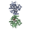
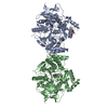
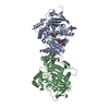
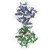
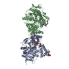
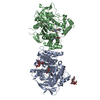
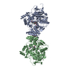
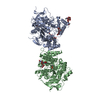
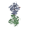
- PDB-7e3h: Crystal structure of human acetylcholinesterase in complex with d... -
+ Open data

- Basic information
| Entry | Database: PDB / ID: 7e3h | ||||||
|---|---|---|---|---|---|---|---|
| Title | Crystal structure of human acetylcholinesterase in complex with donepezil | ||||||
 Components Components | Acetylcholinesterase | ||||||
 Keywords Keywords | HYDROLASE / Human acetylcholinesterase / hAChE / Alzheimer's disease | ||||||
| Function / homology |  Function and homology information Function and homology informationnegative regulation of synaptic transmission, cholinergic / serine hydrolase activity / acetylcholine catabolic process in synaptic cleft / Neurotransmitter clearance / acetylcholine catabolic process / amyloid precursor protein metabolic process / acetylcholinesterase / cholinesterase activity / acetylcholine binding / osteoblast development ...negative regulation of synaptic transmission, cholinergic / serine hydrolase activity / acetylcholine catabolic process in synaptic cleft / Neurotransmitter clearance / acetylcholine catabolic process / amyloid precursor protein metabolic process / acetylcholinesterase / cholinesterase activity / acetylcholine binding / osteoblast development / acetylcholine receptor signaling pathway / acetylcholinesterase activity / Synthesis of PC / basement membrane / Synthesis, secretion, and deacylation of Ghrelin / regulation of receptor recycling / side of membrane / synaptic cleft / collagen binding / laminin binding / synapse assembly / positive regulation of protein secretion / neuromuscular junction / receptor internalization / nervous system development / positive regulation of cold-induced thermogenesis / amyloid-beta binding / retina development in camera-type eye / cell adhesion / hydrolase activity / synapse / perinuclear region of cytoplasm / cell surface / Golgi apparatus / protein homodimerization activity / : / extracellular region / membrane / nucleus / plasma membrane Similarity search - Function | ||||||
| Biological species |  Homo sapiens (human) Homo sapiens (human) | ||||||
| Method |  X-RAY DIFFRACTION / SYNCHROTRON / MOLECULAR REPLACEMENT / Resolution: 2.45 Å X-RAY DIFFRACTION / SYNCHROTRON / MOLECULAR REPLACEMENT / Resolution: 2.45 Å | ||||||
 Authors Authors | Dileep, K.V. / Ihara, K. / Mishima-Tsumagari, C. / Kukimoto-Niino, M. / Yonemochi, M. / Hanada, K. / Shirouzu, M. / Zhang, K.Y.J. | ||||||
 Citation Citation | Journal: Int.J.Biol.Macromol. / Year: 2022 Title: Crystal structure of human acetylcholinesterase in complex with tacrine: Implications for drug discovery Authors: Dileep, K. / Ihara, K. / Mishima-Tsumagari, C. / Kukimoto-Niino, M. / Yonemochi, M. / Hanada, K. / Shirouzu, M. / Zhang, K.Y. | ||||||
| History |
|
- Structure visualization
| Structure viewer | Molecule: MolmilJmol/JSmol |
|---|
- Downloads & links
-Download
| PDBx/mmCIF format | 7e3h.cif.gz | 218.5 KB | Display | PDBx/mmCIF format |
|---|---|---|---|---|
| PDB format | pdb7e3h.ent.gz | 173.1 KB | Display | PDB format |
| PDBx/mmJSON format | 7e3h.json.gz | Tree view | PDBx/mmJSON format | |
| Others |  Other downloads Other downloads |
-Validation report
| Arichive directory | https://data.pdbj.org/pub/pdb/validation_reports/e3/7e3hftp://data.pdbj.org/pub/pdb/validation_reports/e3/7e3h | HTTPS FTP |
|---|
-Related structure data
| Related structure data |  7e3dC 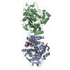 7xn1C  4ey7S S: Starting model for refinement C: citing same article ( |
|---|---|
| Similar structure data |


-Links
 PDBj
PDBj


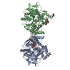
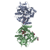
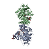
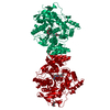
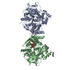
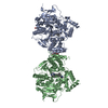
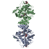
- Assembly
| Deposited unit | 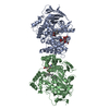
| ||||||||
|---|---|---|---|---|---|---|---|---|---|
| 1 |
| ||||||||
| Unit cell |
|
-Components
| #1: Protein | Mass: 59232.863 Da / Num. of mol.: 2 Source method: isolated from a genetically manipulated source Source: (gene. exp.) Homo sapiens (human) / Gene: ACHE / Cell line (production host): HEK293 / Production host: Homo sapiens (human) / References: UniProt: P22303, acetylcholinesterase#2: Polysaccharide | Source method: isolated from a genetically manipulated source #3: Chemical | 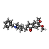  Mass: 379.492 Da / Num. of mol.: 2 / Source method: obtained synthetically / Formula: C24H29NO3 / Feature type: SUBJECT OF INVESTIGATION Mass: 379.492 Da / Num. of mol.: 2 / Source method: obtained synthetically / Formula: C24H29NO3 / Feature type: SUBJECT OF INVESTIGATION#4: Water | ChemComp-HOH / |  Mass: 18.015 Da / Num. of mol.: 57 / Source method: isolated from a natural source / Formula: H2O Mass: 18.015 Da / Num. of mol.: 57 / Source method: isolated from a natural source / Formula: H2OHas ligand of interest | Y | Has protein modification | Y | |
|---|
-Experimental details
-Experiment
| Experiment | Method: X-RAY DIFFRACTION / Number of used crystals: 1 |
|---|
- Sample preparation
| Crystal | Density Matthews: 4.34 Å3/Da / Density % sol: 71.63 % |
|---|---|
| Crystal grow | Temperature: 293.15 K / Method: vapor diffusion, hanging drop / pH: 9 Details: 100 mM Tris HCl buffer pH 9.0, 20 % PEG 3350, 200 mM KNO3 PH range: 8.0 - 9.0 |
-Data collection
| Diffraction | Mean temperature: 100 K / Serial crystal experiment: N | ||||||||||||||||||||||||
|---|---|---|---|---|---|---|---|---|---|---|---|---|---|---|---|---|---|---|---|---|---|---|---|---|---|
| Diffraction source | Source: SYNCHROTRON / Site: SPring-8  / Beamline: BL26B2 / Wavelength: 1 Å / Beamline: BL26B2 / Wavelength: 1 Å | ||||||||||||||||||||||||
| Detector | Type: RAYONIX MX-225 / Detector: CCD / Date: Jul 18, 2019 | ||||||||||||||||||||||||
| Radiation | Protocol: SINGLE WAVELENGTH / Monochromatic (M) / Laue (L): M / Scattering type: x-ray | ||||||||||||||||||||||||
| Radiation wavelength | Wavelength: 1 Å / Relative weight: 1 | ||||||||||||||||||||||||
| Reflection | Resolution: 2.45→47.2 Å / Num. obs: 77020 / % possible obs: 100 % / Redundancy: 22.2 % / CC1/2: 0.999 / Rmerge(I) obs: 0.122 / Net I/σ(I): 23.9 / Num. measured all: 1708002 | ||||||||||||||||||||||||
| Reflection shell | Diffraction-ID: 1
|
- Processing
| Software |
| ||||||||||||||||||||||||||||||||||||||||||||||||||||||||||||||||||||||||||||||||||||||||||||||||||||||||||||||||||||||||||||||||||||||||||||||||||||||||||||||||||||||||||||||
|---|---|---|---|---|---|---|---|---|---|---|---|---|---|---|---|---|---|---|---|---|---|---|---|---|---|---|---|---|---|---|---|---|---|---|---|---|---|---|---|---|---|---|---|---|---|---|---|---|---|---|---|---|---|---|---|---|---|---|---|---|---|---|---|---|---|---|---|---|---|---|---|---|---|---|---|---|---|---|---|---|---|---|---|---|---|---|---|---|---|---|---|---|---|---|---|---|---|---|---|---|---|---|---|---|---|---|---|---|---|---|---|---|---|---|---|---|---|---|---|---|---|---|---|---|---|---|---|---|---|---|---|---|---|---|---|---|---|---|---|---|---|---|---|---|---|---|---|---|---|---|---|---|---|---|---|---|---|---|---|---|---|---|---|---|---|---|---|---|---|---|---|---|---|---|---|
| Refinement | Method to determine structure: MOLECULAR REPLACEMENT Starting model: 4EY7 Resolution: 2.45→47.2 Å / SU ML: 0.27 / Cross valid method: THROUGHOUT / σ(F): 1.33 / Phase error: 24.01 / Stereochemistry target values: ML
| ||||||||||||||||||||||||||||||||||||||||||||||||||||||||||||||||||||||||||||||||||||||||||||||||||||||||||||||||||||||||||||||||||||||||||||||||||||||||||||||||||||||||||||||
| Solvent computation | Shrinkage radii: 0.9 Å / VDW probe radii: 1.11 Å / Solvent model: FLAT BULK SOLVENT MODEL | ||||||||||||||||||||||||||||||||||||||||||||||||||||||||||||||||||||||||||||||||||||||||||||||||||||||||||||||||||||||||||||||||||||||||||||||||||||||||||||||||||||||||||||||
| Displacement parameters | Biso max: 116.2 Å2 / Biso mean: 41.2213 Å2 / Biso min: 21.64 Å2 | ||||||||||||||||||||||||||||||||||||||||||||||||||||||||||||||||||||||||||||||||||||||||||||||||||||||||||||||||||||||||||||||||||||||||||||||||||||||||||||||||||||||||||||||
| Refinement step | Cycle: final / Resolution: 2.45→47.2 Å
| ||||||||||||||||||||||||||||||||||||||||||||||||||||||||||||||||||||||||||||||||||||||||||||||||||||||||||||||||||||||||||||||||||||||||||||||||||||||||||||||||||||||||||||||
| LS refinement shell | Refine-ID: X-RAY DIFFRACTION / Rfactor Rfree error: 0 / Total num. of bins used: 28 / % reflection obs: 100 %
|
