 Movie
Movie Controller


+ Open data

- Basic information
| Entry | Database: PDB / ID: 7e3d | ||||||
|---|---|---|---|---|---|---|---|
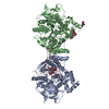
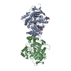
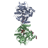
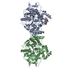
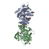
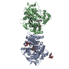
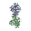
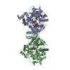
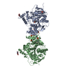
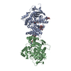
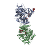
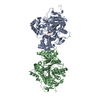
| Title | Crystal structure of human acetylcholinesterase | ||||||
 Components Components | Acetylcholinesterase | ||||||
 Keywords Keywords | HYDROLASE / Human acetylcholinesterase / hAChE / Alzheimer's disease | ||||||
| Function / homology |  Function and homology information Function and homology informationnegative regulation of synaptic transmission, cholinergic / serine hydrolase activity / acetylcholine catabolic process in synaptic cleft / Neurotransmitter clearance / acetylcholine catabolic process / amyloid precursor protein metabolic process / acetylcholinesterase / cholinesterase activity / acetylcholine binding / osteoblast development ...negative regulation of synaptic transmission, cholinergic / serine hydrolase activity / acetylcholine catabolic process in synaptic cleft / Neurotransmitter clearance / acetylcholine catabolic process / amyloid precursor protein metabolic process / acetylcholinesterase / cholinesterase activity / acetylcholine binding / osteoblast development / acetylcholine receptor signaling pathway / acetylcholinesterase activity / Synthesis of PC / basement membrane / Synthesis, secretion, and deacylation of Ghrelin / regulation of receptor recycling / side of membrane / synaptic cleft / collagen binding / laminin binding / synapse assembly / positive regulation of protein secretion / neuromuscular junction / receptor internalization / nervous system development / positive regulation of cold-induced thermogenesis / amyloid-beta binding / retina development in camera-type eye / cell adhesion / hydrolase activity / synapse / perinuclear region of cytoplasm / cell surface / Golgi apparatus / protein homodimerization activity / : / extracellular region / membrane / nucleus / plasma membrane Similarity search - Function | ||||||
| Biological species |  Homo sapiens (human) Homo sapiens (human) | ||||||
| Method |  X-RAY DIFFRACTION / SYNCHROTRON / MOLECULAR REPLACEMENT / Resolution: 2.5 Å X-RAY DIFFRACTION / SYNCHROTRON / MOLECULAR REPLACEMENT / Resolution: 2.5 Å | ||||||
 Authors Authors | Dileep, K.V. / Ihara, K. / Mishima-Tsumagari, C. / Kukimoto-Niino, M. / Yonemochi, M. / Hanada, K. / Shirouzu, M. / Zhang, K.Y.J. | ||||||
 Citation Citation | Journal: Int.J.Biol.Macromol. / Year: 2022 Title: Crystal structure of human acetylcholinesterase in complex with tacrine: Implications for drug discovery Authors: Dileep, K. / Ihara, K. / Mishima-Tsumagari, C. / Kukimoto-Niino, M. / Yonemochi, M. / Hanada, K. / Shirouzu, M. / Zhang, K.Y. | ||||||
| History |
|
- Structure visualization
| Structure viewer | Molecule: MolmilJmol/JSmol |
|---|
- Downloads & links
-Download
| PDBx/mmCIF format | 7e3d.cif.gz | 222.4 KB | Display | PDBx/mmCIF format |
|---|---|---|---|---|
| PDB format | pdb7e3d.ent.gz | 175.3 KB | Display | PDB format |
| PDBx/mmJSON format | 7e3d.json.gz | Tree view | PDBx/mmJSON format | |
| Others |  Other downloads Other downloads |
-Validation report
| Arichive directory | https://data.pdbj.org/pub/pdb/validation_reports/e3/7e3dftp://data.pdbj.org/pub/pdb/validation_reports/e3/7e3d | HTTPS FTP |
|---|
-Related structure data
| Related structure data | 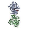 7e3hC 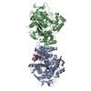 7xn1C  4ey4S S: Starting model for refinement C: citing same article ( |
|---|---|
| Similar structure data |


-Links
 PDBj
PDBj


- Assembly
| Deposited unit | 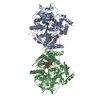
| ||||||||||||||||||||||||||||||||||||||||||||||||||||||||||||||||||||||||||||||||||||||||||||||||||||||||
|---|---|---|---|---|---|---|---|---|---|---|---|---|---|---|---|---|---|---|---|---|---|---|---|---|---|---|---|---|---|---|---|---|---|---|---|---|---|---|---|---|---|---|---|---|---|---|---|---|---|---|---|---|---|---|---|---|---|---|---|---|---|---|---|---|---|---|---|---|---|---|---|---|---|---|---|---|---|---|---|---|---|---|---|---|---|---|---|---|---|---|---|---|---|---|---|---|---|---|---|---|---|---|---|---|---|
| 1 |
| ||||||||||||||||||||||||||||||||||||||||||||||||||||||||||||||||||||||||||||||||||||||||||||||||||||||||
| Unit cell |
| ||||||||||||||||||||||||||||||||||||||||||||||||||||||||||||||||||||||||||||||||||||||||||||||||||||||||
| Noncrystallographic symmetry (NCS) | NCS domain:
NCS domain segments:
|