 Movie
Movie Controller


+ Open data

- Basic information
| Entry | Database: PDB / ID: 7e1b | ||||||
|---|---|---|---|---|---|---|---|
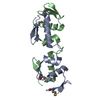
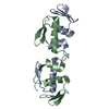
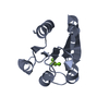
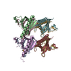
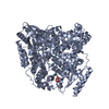
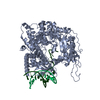
| Title | Crystal structure of VbrR-DNA complex | ||||||
 Components Components |
| ||||||
 Keywords Keywords | DNA BINDING PROTEIN / Response regulator | ||||||
| Function / homology |  Function and homology information Function and homology informationphosphorelay response regulator activity / protein-DNA complex / transcription cis-regulatory region binding / regulation of DNA-templated transcription / cytosol Similarity search - Function | ||||||
| Biological species |   Vibrio parahaemolyticus (bacteria) Vibrio parahaemolyticus (bacteria) | ||||||
| Method |  X-RAY DIFFRACTION / SYNCHROTRON / SAD / Resolution: 4.587 Å X-RAY DIFFRACTION / SYNCHROTRON / SAD / Resolution: 4.587 Å | ||||||
 Authors Authors | Hong, S. / Zhang, X. / Zhang, P. | ||||||
| Funding support |  China, 1items China, 1items
| ||||||
 Citation Citation | Journal: Acta Biochim.Biophys.Sin. / Year: 2023 Title: Structural basis of phosphorylation-induced activation of the response regulator VbrR. Authors: Hong, S. / Guo, J. / Zhang, X. / Zhou, X. / Zhang, P. / Yu, F. | ||||||
| History |
|
- Structure visualization
| Structure viewer | Molecule: MolmilJmol/JSmol |
|---|
- Downloads & links
-Download
| PDBx/mmCIF format | 7e1b.cif.gz | 378.1 KB | Display | PDBx/mmCIF format |
|---|---|---|---|---|
| PDB format | pdb7e1b.ent.gz | 304.3 KB | Display | PDB format |
| PDBx/mmJSON format | 7e1b.json.gz | Tree view | PDBx/mmJSON format | |
| Others |  Other downloads Other downloads |
-Validation report
| Arichive directory | https://data.pdbj.org/pub/pdb/validation_reports/e1/7e1bftp://data.pdbj.org/pub/pdb/validation_reports/e1/7e1b | HTTPS FTP |
|---|
-Related structure data


-Links
 PDBj
PDBj


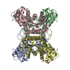
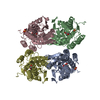
- Assembly
| Deposited unit | 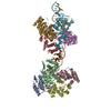
| ||||||||
|---|---|---|---|---|---|---|---|---|---|
| 1 | 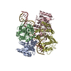
| ||||||||
| 2 | 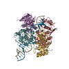
| ||||||||
| Unit cell |
|
-Components
| #1: Protein | Mass: 24906.615 Da / Num. of mol.: 8 Source method: isolated from a genetically manipulated source Source: (gene. exp.) Vibrio parahaemolyticus (bacteria)Gene: C9I78_18460, CA163_10520, CGH73_12770, CGI34_20580, CGI42_14750, D5E78_24805, F0L89_03250, F0L99_22745, WR32_19050 Production host: #2: DNA chain | Mass: 7863.088 Da / Num. of mol.: 2 Source method: isolated from a genetically manipulated source Source: (gene. exp.) Vibrio parahaemolyticus (bacteria) / Production host: Vibrio parahaemolyticus (bacteria)#3: DNA chain | Mass: 8108.279 Da / Num. of mol.: 2 Source method: isolated from a genetically manipulated source Source: (gene. exp.) Vibrio parahaemolyticus (bacteria) / Production host: Vibrio parahaemolyticus (bacteria) |
|---|
-Experimental details
-Experiment
| Experiment | Method: X-RAY DIFFRACTION / Number of used crystals: 1 |
|---|
- Sample preparation
| Crystal | Density Matthews: 4.36 Å3/Da / Density % sol: 71.79 % |
|---|---|
| Crystal grow | Temperature: 293 K / Method: vapor diffusion, sitting drop / Details: 0.2 M NaCl, 0.1 M MES pH 6.6, and 20% PEG4000 |
-Data collection
| Diffraction | Mean temperature: 100 K / Serial crystal experiment: N | |||||||||||||||||||||||||||||||||||||||||||||||||||||||||||||||||||||||||||||||||||||||||||||||||||
|---|---|---|---|---|---|---|---|---|---|---|---|---|---|---|---|---|---|---|---|---|---|---|---|---|---|---|---|---|---|---|---|---|---|---|---|---|---|---|---|---|---|---|---|---|---|---|---|---|---|---|---|---|---|---|---|---|---|---|---|---|---|---|---|---|---|---|---|---|---|---|---|---|---|---|---|---|---|---|---|---|---|---|---|---|---|---|---|---|---|---|---|---|---|---|---|---|---|---|---|---|
| Diffraction source | Source: SYNCHROTRON / Site: SSRF / Beamline: BL17U1 / Wavelength: 0.97818 Å | |||||||||||||||||||||||||||||||||||||||||||||||||||||||||||||||||||||||||||||||||||||||||||||||||||
| Detector | Type: ADSC QUANTUM 315r / Detector: CCD / Date: Apr 11, 2019 | |||||||||||||||||||||||||||||||||||||||||||||||||||||||||||||||||||||||||||||||||||||||||||||||||||
| Radiation | Protocol: SINGLE WAVELENGTH / Monochromatic (M) / Laue (L): M / Scattering type: x-ray | |||||||||||||||||||||||||||||||||||||||||||||||||||||||||||||||||||||||||||||||||||||||||||||||||||
| Radiation wavelength | Wavelength: 0.97818 Å / Relative weight: 1 | |||||||||||||||||||||||||||||||||||||||||||||||||||||||||||||||||||||||||||||||||||||||||||||||||||
| Reflection twin | Operator: -h-k-l,l,k / Fraction: 0.45 | |||||||||||||||||||||||||||||||||||||||||||||||||||||||||||||||||||||||||||||||||||||||||||||||||||
| Reflection | Resolution: 4.587→50 Å / Num. obs: 15995 / % possible obs: 93.7 % / Redundancy: 3.2 % / Rmerge(I) obs: 0.065 / Rpim(I) all: 0.042 / Rrim(I) all: 0.078 / Χ2: 0.843 / Net I/σ(I): 7.2 | |||||||||||||||||||||||||||||||||||||||||||||||||||||||||||||||||||||||||||||||||||||||||||||||||||
| Reflection shell | Diffraction-ID: 1
|
- Processing
| Software |
| ||||||||||||||||||||||||||||||||||||||||||||||||||||||||||||||||||||||||
|---|---|---|---|---|---|---|---|---|---|---|---|---|---|---|---|---|---|---|---|---|---|---|---|---|---|---|---|---|---|---|---|---|---|---|---|---|---|---|---|---|---|---|---|---|---|---|---|---|---|---|---|---|---|---|---|---|---|---|---|---|---|---|---|---|---|---|---|---|---|---|---|---|---|
| Refinement | Method to determine structure: SAD / Resolution: 4.587→46.006 Å / Cross valid method: THROUGHOUT / σ(F): 16.35 / Phase error: 34 / Stereochemistry target values: TWIN_LSQ_F
| ||||||||||||||||||||||||||||||||||||||||||||||||||||||||||||||||||||||||
| Solvent computation | Shrinkage radii: 0.9 Å / VDW probe radii: 1.11 Å / Solvent model: FLAT BULK SOLVENT MODEL | ||||||||||||||||||||||||||||||||||||||||||||||||||||||||||||||||||||||||
| Displacement parameters | Biso max: 185.28 Å2 / Biso mean: 140.2032 Å2 / Biso min: 80.24 Å2 | ||||||||||||||||||||||||||||||||||||||||||||||||||||||||||||||||||||||||
| Refinement step | Cycle: final / Resolution: 4.587→46.006 Å
| ||||||||||||||||||||||||||||||||||||||||||||||||||||||||||||||||||||||||
| LS refinement shell | Refine-ID: X-RAY DIFFRACTION / Rfactor Rfree error: 0
|
