 Movie
Movie Controller

[English] 日本語
 Yorodumi
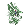
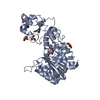
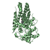
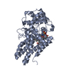
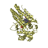
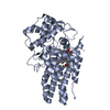
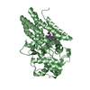
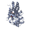
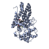
- PDB-7e0u: Crystal Structure of Human Indoleamine 2,3-dioxygenagse 1 (hIDO1)... -
+ Open data

- Basic information
| Entry | Database: PDB / ID: 7e0u | ||||||
|---|---|---|---|---|---|---|---|
| Title | Crystal Structure of Human Indoleamine 2,3-dioxygenagse 1 (hIDO1) Complexed with 6-Bromo-N-(((1S,2S)-2-chlorocyclohexyl)methyl)-1H-indazol-4-amine (39) | ||||||
 Components Components | Indoleamine 2,3-dioxygenase 1 | ||||||
 Keywords Keywords | OXIDOREDUCTASE / Indoleamine 2 / 3-dioxygenase 1 | ||||||
| Function / homology |  Function and homology information Function and homology information indoleamine 2,3-dioxygenase / positive regulation of chronic inflammatory response / kynurenic acid biosynthetic process / smooth muscle contractile fiber / indoleamine 2,3-dioxygenase activity / 'de novo' NAD+ biosynthetic process from L-tryptophan / L-tryptophan 2,3-dioxygenase activity / positive regulation of T cell tolerance induction / : / stereocilium bundle ... indoleamine 2,3-dioxygenase / positive regulation of chronic inflammatory response / kynurenic acid biosynthetic process / smooth muscle contractile fiber / indoleamine 2,3-dioxygenase activity / 'de novo' NAD+ biosynthetic process from L-tryptophan / L-tryptophan 2,3-dioxygenase activity / positive regulation of T cell tolerance induction / : / stereocilium bundle / positive regulation of type 2 immune response / L-tryptophan catabolic process / Tryptophan catabolism / negative regulation of T cell apoptotic process / positive regulation of T cell apoptotic process / quinolinate biosynthetic process / negative regulation of interleukin-10 production / T cell proliferation / multicellular organismal response to stress / negative regulation of T cell proliferation / swimming behavior / positive regulation of interleukin-12 production / female pregnancy / response to lipopolysaccharide / electron transfer activity / inflammatory response / heme binding / metal ion binding / cytoplasm / cytosol Similarity search - Function | ||||||
| Biological species |  Homo sapiens (human) Homo sapiens (human) | ||||||
| Method |  X-RAY DIFFRACTION / SYNCHROTRON / MOLECULAR REPLACEMENT / Resolution: 2.278 Å X-RAY DIFFRACTION / SYNCHROTRON / MOLECULAR REPLACEMENT / Resolution: 2.278 Å | ||||||
 Authors Authors | Li, G.-B. / Ning, X.-L. | ||||||
| Funding support |  China, 1items China, 1items
| ||||||
 Citation Citation | Journal: J.Med.Chem. / Year: 2021 Title: X-ray Structure-Guided Discovery of a Potent, Orally Bioavailable, Dual Human Indoleamine/Tryptophan 2,3-Dioxygenase (hIDO/hTDO) Inhibitor That Shows Activity in a Mouse Model of Parkinson's Disease. Authors: Ning, X.L. / Li, Y.Z. / Huo, C. / Deng, J. / Gao, C. / Zhu, K.R. / Wang, M. / Wu, Y.X. / Yu, J.L. / Ren, Y.L. / Luo, Z.Y. / Li, G. / Chen, Y. / Wang, S.Y. / Peng, C. / Yang, L.L. / Wang, Z.Y. ...Authors: Ning, X.L. / Li, Y.Z. / Huo, C. / Deng, J. / Gao, C. / Zhu, K.R. / Wang, M. / Wu, Y.X. / Yu, J.L. / Ren, Y.L. / Luo, Z.Y. / Li, G. / Chen, Y. / Wang, S.Y. / Peng, C. / Yang, L.L. / Wang, Z.Y. / Wu, Y. / Qian, S. / Li, G.B. | ||||||
| History |
|
- Structure visualization
| Structure viewer | Molecule: MolmilJmol/JSmol |
|---|
- Downloads & links
-Download
| PDBx/mmCIF format | 7e0u.cif.gz | 169.3 KB | Display | PDBx/mmCIF format |
|---|---|---|---|---|
| PDB format | pdb7e0u.ent.gz | 131.8 KB | Display | PDB format |
| PDBx/mmJSON format | 7e0u.json.gz | Tree view | PDBx/mmJSON format | |
| Others |  Other downloads Other downloads |
-Validation report
| Arichive directory | https://data.pdbj.org/pub/pdb/validation_reports/e0/7e0uftp://data.pdbj.org/pub/pdb/validation_reports/e0/7e0u | HTTPS FTP |
|---|
-Related structure data
| Related structure data | 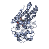 7e0oC  7e0pC  7e0qC  7e0sC  7e0tC  5etwS S: Starting model for refinement C: citing same article ( |
|---|---|
| Similar structure data |
-Links
 PDBj
PDBj- Assembly
| Deposited unit | 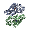
| ||||||||
|---|---|---|---|---|---|---|---|---|---|
| 1 | 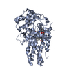
| ||||||||
| 2 | 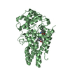
| ||||||||
| Unit cell |
|
-Components
| #1: Protein | Mass: 44126.711 Da / Num. of mol.: 2 Source method: isolated from a genetically manipulated source Source: (gene. exp.) Homo sapiens (human) / Gene: IDO1, IDO, INDO / Production host:  #2: Chemical |   Mass: 616.487 Da / Num. of mol.: 2 / Source method: obtained synthetically / Formula: C34H32FeN4O4 / Feature type: SUBJECT OF INVESTIGATION Mass: 616.487 Da / Num. of mol.: 2 / Source method: obtained synthetically / Formula: C34H32FeN4O4 / Feature type: SUBJECT OF INVESTIGATION#3: Chemical | 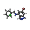  Mass: 342.662 Da / Num. of mol.: 2 / Source method: obtained synthetically / Formula: C14H17BrClN3 / Feature type: SUBJECT OF INVESTIGATION Mass: 342.662 Da / Num. of mol.: 2 / Source method: obtained synthetically / Formula: C14H17BrClN3 / Feature type: SUBJECT OF INVESTIGATION#4: Water | ChemComp-HOH / |  Mass: 18.015 Da / Num. of mol.: 108 / Source method: isolated from a natural source / Formula: H2O Mass: 18.015 Da / Num. of mol.: 108 / Source method: isolated from a natural source / Formula: H2OHas ligand of interest | Y | Has protein modification | Y | |
|---|
-Experimental details
-Experiment
| Experiment | Method: X-RAY DIFFRACTION / Number of used crystals: 1 |
|---|
- Sample preparation
| Crystal | Density Matthews: 2.84 Å3/Da / Density % sol: 56.67 % |
|---|---|
| Crystal grow | Temperature: 293 K / Method: vapor diffusion, hanging drop / Details: 15% to 23% PEG 8000, 0.2M ammonium acetate |
-Data collection
| Diffraction | Mean temperature: 195 K / Serial crystal experiment: N | ||||||||||||||||||||||||||||||
|---|---|---|---|---|---|---|---|---|---|---|---|---|---|---|---|---|---|---|---|---|---|---|---|---|---|---|---|---|---|---|---|
| Diffraction source | Source: SYNCHROTRON / Site: SSRF / Beamline: BL19U1 / Wavelength: 0.97918 Å | ||||||||||||||||||||||||||||||
| Detector | Type: DECTRIS PILATUS3 X CdTe 1M / Detector: PIXEL / Date: Oct 29, 2020 | ||||||||||||||||||||||||||||||
| Radiation | Protocol: SINGLE WAVELENGTH / Monochromatic (M) / Laue (L): M / Scattering type: x-ray | ||||||||||||||||||||||||||||||
| Radiation wavelength | Wavelength: 0.97918 Å / Relative weight: 1 | ||||||||||||||||||||||||||||||
| Reflection | Resolution: 2.278→74.453 Å / Num. obs: 46598 / % possible obs: 99.8 % / Redundancy: 12.9 % / Biso Wilson estimate: 53.42 Å2 / CC1/2: 0.999 / Rmerge(I) obs: 0.078 / Rpim(I) all: 0.023 / Rrim(I) all: 0.081 / Net I/σ(I): 19 | ||||||||||||||||||||||||||||||
| Reflection shell | Diffraction-ID: 1
|
- Processing
| Software |
| ||||||||||||||||||||||||||||||||||||||||||||||||||||||||||||||||||||||||||||||||||||||||||
|---|---|---|---|---|---|---|---|---|---|---|---|---|---|---|---|---|---|---|---|---|---|---|---|---|---|---|---|---|---|---|---|---|---|---|---|---|---|---|---|---|---|---|---|---|---|---|---|---|---|---|---|---|---|---|---|---|---|---|---|---|---|---|---|---|---|---|---|---|---|---|---|---|---|---|---|---|---|---|---|---|---|---|---|---|---|---|---|---|---|---|---|
| Refinement | Method to determine structure: MOLECULAR REPLACEMENT Starting model: 5etw Resolution: 2.278→74.453 Å / SU ML: 0.28 / Cross valid method: THROUGHOUT / σ(F): 1.35 / Phase error: 28.07 / Stereochemistry target values: ML
| ||||||||||||||||||||||||||||||||||||||||||||||||||||||||||||||||||||||||||||||||||||||||||
| Solvent computation | Shrinkage radii: 0.9 Å / VDW probe radii: 1.11 Å / Solvent model: FLAT BULK SOLVENT MODEL | ||||||||||||||||||||||||||||||||||||||||||||||||||||||||||||||||||||||||||||||||||||||||||
| Displacement parameters | Biso max: 128.5 Å2 / Biso mean: 63.9333 Å2 / Biso min: 34.72 Å2 | ||||||||||||||||||||||||||||||||||||||||||||||||||||||||||||||||||||||||||||||||||||||||||
| Refinement step | Cycle: final / Resolution: 2.278→74.453 Å
| ||||||||||||||||||||||||||||||||||||||||||||||||||||||||||||||||||||||||||||||||||||||||||
| Refine LS restraints |
| ||||||||||||||||||||||||||||||||||||||||||||||||||||||||||||||||||||||||||||||||||||||||||
| LS refinement shell | Refine-ID: X-RAY DIFFRACTION / Rfactor Rfree error: 0
|
