 Movie
Movie Controller


+ Open data

- Basic information
| Entry | Database: PDB / ID: 7dya | ||||||
|---|---|---|---|---|---|---|---|
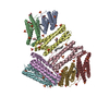
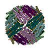
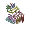
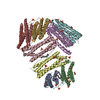
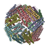
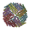
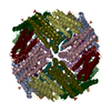
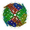
| Title | Crystal structure of TmFtn with calcium ions | ||||||
 Components Components | Ferritin | ||||||
 Keywords Keywords | PROTEIN BINDING / Thermotoga maritima ferritin / Calcium ions | ||||||
| Function / homology |  Function and homology information Function and homology informationbacterial non-heme ferritin / ferroxidase activity / ferric iron binding / iron ion transport / ferrous iron binding / intracellular iron ion homeostasis / identical protein binding / cytoplasm / cytosol Similarity search - Function | ||||||
| Biological species |   Thermotoga maritima (bacteria) Thermotoga maritima (bacteria) | ||||||
| Method |  X-RAY DIFFRACTION / SYNCHROTRON / MOLECULAR REPLACEMENT / Resolution: 2.197 Å X-RAY DIFFRACTION / SYNCHROTRON / MOLECULAR REPLACEMENT / Resolution: 2.197 Å | ||||||
 Authors Authors | Zhang, X. / Zhao, G. | ||||||
| Funding support |  China, 1items China, 1items
| ||||||
 Citation Citation | Journal: Nat Commun / Year: 2021 Title: Protein interface redesign facilitates the transformation of nanocage building blocks to 1D and 2D nanomaterials. Authors: Zhang, X. / Liu, Y. / Zheng, B. / Zang, J. / Lv, C. / Zhang, T. / Wang, H. / Zhao, G. | ||||||
| History |
|
- Structure visualization
| Structure viewer | Molecule: MolmilJmol/JSmol |
|---|
- Downloads & links
-Download
| PDBx/mmCIF format | 7dya.cif.gz | 299.8 KB | Display | PDBx/mmCIF format |
|---|---|---|---|---|
| PDB format | pdb7dya.ent.gz | 244.3 KB | Display | PDB format |
| PDBx/mmJSON format | 7dya.json.gz | Tree view | PDBx/mmJSON format | |
| Others |  Other downloads Other downloads |
-Validation report
| Arichive directory | https://data.pdbj.org/pub/pdb/validation_reports/dy/7dyaftp://data.pdbj.org/pub/pdb/validation_reports/dy/7dya | HTTPS FTP |
|---|
-Related structure data
| Related structure data |  7dy8C 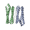 7dy9C  7dybC  1vlgS S: Starting model for refinement C: citing same article ( |
|---|---|
| Similar structure data |


-Links
 PDBj
PDBj


- Assembly
| Deposited unit | 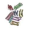
| ||||||||
|---|---|---|---|---|---|---|---|---|---|
| 1 | 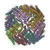
| ||||||||
| Unit cell |
| ||||||||
| Components on special symmetry positions |
|
-Components
| #1: Protein | Mass: 19401.840 Da / Num. of mol.: 8 Source method: isolated from a genetically manipulated source Source: (gene. exp.) Thermotoga maritima (strain ATCC 43589 / MSB8 / DSM 3109 / JCM 10099) (bacteria)Strain: ATCC 43589 / MSB8 / DSM 3109 / JCM 10099 / Gene: TM_1128 / Production host: #2: Chemical | ChemComp-FE /   Mass: 55.845 Da / Num. of mol.: 16 / Source method: obtained synthetically / Formula: Fe Mass: 55.845 Da / Num. of mol.: 16 / Source method: obtained synthetically / Formula: Fe#3: Chemical | ChemComp-CA /   Mass: 40.078 Da / Num. of mol.: 9 / Source method: obtained synthetically / Formula: Ca / Feature type: SUBJECT OF INVESTIGATION Mass: 40.078 Da / Num. of mol.: 9 / Source method: obtained synthetically / Formula: Ca / Feature type: SUBJECT OF INVESTIGATION#4: Water | ChemComp-HOH / |  Mass: 18.015 Da / Num. of mol.: 1019 / Source method: isolated from a natural source / Formula: H2O Mass: 18.015 Da / Num. of mol.: 1019 / Source method: isolated from a natural source / Formula: H2OHas ligand of interest | Y | |
|---|
-Experimental details
-Experiment
| Experiment | Method: X-RAY DIFFRACTION / Number of used crystals: 1 |
|---|
- Sample preparation
| Crystal | Density Matthews: 3.41 Å3/Da / Density % sol: 63.93 % |
|---|---|
| Crystal grow | Temperature: 293 K / Method: vapor diffusion / pH: 4.6 Details: 14% 2-Propanol, 70 mM Sodium acetate/ HCl pH 4.6, 140 mM Calcium chloride, 30% Glycerol |
-Data collection
| Diffraction | Mean temperature: 100 K / Serial crystal experiment: N |
|---|---|
| Diffraction source | Source: SYNCHROTRON / Site: NFPSS / Beamline: BL18U / Wavelength: 0.979 Å |
| Detector | Type: RAYONIX MX-225 / Detector: CCD / Date: Jul 17, 2020 |
| Radiation | Protocol: SINGLE WAVELENGTH / Monochromatic (M) / Laue (L): M / Scattering type: x-ray |
| Radiation wavelength | Wavelength: 0.979 Å / Relative weight: 1 |
| Reflection | Resolution: 2.197→38.584 Å / Num. obs: 107433 / % possible obs: 99.92 % / Redundancy: 6.8 % / CC1/2: 0.992 / Net I/σ(I): 5.97 |
| Reflection shell | Resolution: 2.197→2.197 Å / Num. unique obs: 10652 / CC1/2: 0.979 |
- Processing
| Software |
| |||||||||||||||||||||||||||||||||||||||||||||||||||||||||||||||||||||||||||
|---|---|---|---|---|---|---|---|---|---|---|---|---|---|---|---|---|---|---|---|---|---|---|---|---|---|---|---|---|---|---|---|---|---|---|---|---|---|---|---|---|---|---|---|---|---|---|---|---|---|---|---|---|---|---|---|---|---|---|---|---|---|---|---|---|---|---|---|---|---|---|---|---|---|---|---|---|
| Refinement | Method to determine structure: MOLECULAR REPLACEMENT Starting model: 1VLG Resolution: 2.197→38.584 Å / SU ML: 0.17 / Cross valid method: THROUGHOUT / σ(F): 1.36 / Phase error: 19.64 / Stereochemistry target values: ML
| |||||||||||||||||||||||||||||||||||||||||||||||||||||||||||||||||||||||||||
| Solvent computation | Shrinkage radii: 0.9 Å / VDW probe radii: 1.11 Å / Solvent model: FLAT BULK SOLVENT MODEL | |||||||||||||||||||||||||||||||||||||||||||||||||||||||||||||||||||||||||||
| Displacement parameters | Biso max: 97.79 Å2 / Biso mean: 38.2521 Å2 / Biso min: 19.61 Å2 | |||||||||||||||||||||||||||||||||||||||||||||||||||||||||||||||||||||||||||
| Refinement step | Cycle: final / Resolution: 2.197→38.584 Å
| |||||||||||||||||||||||||||||||||||||||||||||||||||||||||||||||||||||||||||
| LS refinement shell | Refine-ID: X-RAY DIFFRACTION / Rfactor Rfree error: 0 / % reflection obs: 100 %
|
