 Movie
Movie Controller


+ Open data

- Basic information
| Entry | Database: PDB / ID: 7dxj | |||||||||||||||
|---|---|---|---|---|---|---|---|---|---|---|---|---|---|---|---|---|
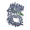
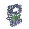
| Title | Human 46QHuntingtin-HAP40 complex structure | |||||||||||||||
 Components Components |
| |||||||||||||||
 Keywords Keywords | STRUCTURAL PROTEIN / Huntingtin / 46Q / HAP40 | |||||||||||||||
| Function / homology |  Function and homology information Function and homology informationvesicle cytoskeletal trafficking / : / positive regulation of CAMKK-AMPK signaling cascade / vocal learning / negative regulation of proteasomal protein catabolic process / regulation of CAMKK-AMPK signaling cascade / positive regulation of mitophagy / profilin binding / positive regulation of cilium assembly / retrograde vesicle-mediated transport, Golgi to endoplasmic reticulum ...vesicle cytoskeletal trafficking / : / positive regulation of CAMKK-AMPK signaling cascade / vocal learning / negative regulation of proteasomal protein catabolic process / regulation of CAMKK-AMPK signaling cascade / positive regulation of mitophagy / profilin binding / positive regulation of cilium assembly / retrograde vesicle-mediated transport, Golgi to endoplasmic reticulum / vesicle transport along microtubule / positive regulation of aggrephagy / positive regulation of lipophagy / establishment of mitotic spindle orientation / Golgi organization / dynein intermediate chain binding / synaptic vesicle transport / dynactin binding / Regulation of MECP2 expression and activity / postsynaptic cytosol / beta-tubulin binding / presynaptic cytosol / phosphoprotein phosphatase activity / heat shock protein binding / inclusion body / neurogenesis / autophagosome / cytoplasmic vesicle membrane / negative regulation of extrinsic apoptotic signaling pathway / central nervous system development / centriole / protein destabilization / kinase binding / p53 binding / late endosome / cytoplasmic vesicle / transmembrane transporter binding / early endosome / nuclear body / positive regulation of apoptotic process / axon / apoptotic process / dendrite / perinuclear region of cytoplasm / Golgi apparatus / endoplasmic reticulum / protein-containing complex / nucleoplasm / identical protein binding / nucleus / cytoplasm / cytosol Similarity search - Function | |||||||||||||||
| Biological species |  Homo sapiens (human) Homo sapiens (human)synthetic construct (others) | |||||||||||||||
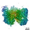
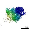
| Method | ELECTRON MICROSCOPY / single particle reconstruction / cryo EM / Resolution: 3.6 Å | |||||||||||||||
 Authors Authors | Guo, Q. / Fernandez-Busnadiego, R. | |||||||||||||||
| Funding support |  Germany, European Union, 4items Germany, European Union, 4items
| |||||||||||||||
 Citation Citation | Journal: Structure / Year: 2021 Title: Pathological polyQ expansion does not alter the conformation of the Huntingtin-HAP40 complex. Authors: Bin Huang / Qiang Guo / Marie L Niedermeier / Jingdong Cheng / Tatjana Engler / Melanie Maurer / Alexander Pautsch / Wolfgang Baumeister / Florian Stengel / Stefan Kochanek / Rubén Fernández-Busnadiego /  Abstract: The abnormal amplification of a CAG repeat in the gene coding for huntingtin (HTT) leads to Huntington's disease (HD). At the protein level, this translates into the expansion of a polyglutamine ...The abnormal amplification of a CAG repeat in the gene coding for huntingtin (HTT) leads to Huntington's disease (HD). At the protein level, this translates into the expansion of a polyglutamine (polyQ) stretch located at the HTT N terminus, which renders HTT aggregation prone by unknown mechanisms. Here we investigated the effects of polyQ expansion on HTT in a complex with its stabilizing interaction partner huntingtin-associated protein 40 (HAP40). Surprisingly, our comprehensive biophysical, crosslinking mass spectrometry and cryo-EM experiments revealed no major differences in the conformation of HTT-HAP40 complexes of various polyQ length, including 17QHTT-HAP40 (wild type), 46QHTT-HAP40 (typical polyQ length in HD patients), and 128QHTT-HAP40 (extreme polyQ length). Thus, HTT polyQ expansion does not alter the global conformation of HTT when associated with HAP40. | |||||||||||||||
| History |
|
- Structure visualization
| Movie |
Movie viewer |
|---|---|
| Structure viewer | Molecule: MolmilJmol/JSmol |
- Downloads & links
-Download
| PDBx/mmCIF format | 7dxj.cif.gz | 479 KB | Display | PDBx/mmCIF format |
|---|---|---|---|---|
| PDB format | pdb7dxj.ent.gz | 372.1 KB | Display | PDB format |
| PDBx/mmJSON format | 7dxj.json.gz | Tree view | PDBx/mmJSON format | |
| Others |  Other downloads Other downloads |
-Validation report
| Arichive directory | https://data.pdbj.org/pub/pdb/validation_reports/dx/7dxjftp://data.pdbj.org/pub/pdb/validation_reports/dx/7dxj | HTTPS FTP |
|---|
-Related structure data
| Related structure data |  30911MC  7dxkC M: map data used to model this data C: citing same article ( |
|---|---|
| Similar structure data | |
| EM raw data | EMPIAR-10667 (Title: Human 46QHuntingtin-HAP40 complex structure / Data size: 59.2 Data #1: Aligned micrographs of Human 46QHuntingtin-HAP40 sample [micrographs - single frame]) |


-Links
 PDBj
PDBj


- Assembly
| Deposited unit | 
|
|---|---|
| 1 |
|
-Components
| #1: Protein | Mass: 351191.312 Da / Num. of mol.: 1 Source method: isolated from a genetically manipulated source Details: 46Q-Huntingtin Source: (gene. exp.) Homo sapiens (human), (gene. exp.) synthetic construct (others)Gene: HTT, HD, IT15 / Production host:  |
|---|---|
| #2: Protein | Mass: 39141.879 Da / Num. of mol.: 1 Source method: isolated from a genetically manipulated source Source: (gene. exp.) Homo sapiens (human) / Gene: F8A1, F8A2, F8A3 / Production host: |
| Has protein modification | Y |
-Experimental details
-Experiment
| Experiment | Method: ELECTRON MICROSCOPY |
|---|---|
| EM experiment | Aggregation state: PARTICLE / 3D reconstruction method: single particle reconstruction |
- Sample preparation
| Component | Name: Human 46QHuntingtin-HAP40 complex structure / Type: COMPLEX / Entity ID: all / Source: RECOMBINANT |
|---|---|
| Source (natural) | Organism: Homo sapiens (human) |
| Source (recombinant) | Organism: |
| Buffer solution | pH: 7 |
| Specimen | Embedding applied: NO / Shadowing applied: NO / Staining applied: NO / Vitrification applied: YES |
| Specimen support | Grid material: GOLD / Grid type: Quantifoil |
| Vitrification | Cryogen name: ETHANE-PROPANE |
- Electron microscopy imaging
| Experimental equipment |  Model: Titan Krios / Image courtesy: FEI Company |
|---|---|
| Microscopy | Model: FEI TITAN KRIOS |
| Electron gun | Electron source:  FIELD EMISSION GUN / Accelerating voltage: 300 kV / Illumination mode: FLOOD BEAM FIELD EMISSION GUN / Accelerating voltage: 300 kV / Illumination mode: FLOOD BEAM |
| Electron lens | Mode: BRIGHT FIELD |
| Image recording | Electron dose: 60 e/Å2 / Film or detector model: GATAN K2 SUMMIT (4k x 4k) |
- Processing
| EM software |
| |||||||||
|---|---|---|---|---|---|---|---|---|---|---|
| CTF correction | Type: PHASE FLIPPING AND AMPLITUDE CORRECTION | |||||||||
| 3D reconstruction | Resolution: 3.6 Å / Resolution method: FSC 0.143 CUT-OFF / Num. of particles: 192473 / Symmetry type: POINT |
