 Movie
Movie Controller

[English] 日本語
 Yorodumi
- PDB-7dwq: Photosystem I from a chlorophyll d-containing cyanobacterium Acar... -
+ Open data

- Basic information
| Entry | Database: PDB / ID: 7dwq | |||||||||||||||||||||||||||
|---|---|---|---|---|---|---|---|---|---|---|---|---|---|---|---|---|---|---|---|---|---|---|---|---|---|---|---|---|
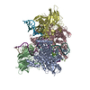
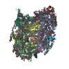
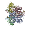
| Title | Photosystem I from a chlorophyll d-containing cyanobacterium Acaryochloris marina | |||||||||||||||||||||||||||
 Components Components |
| |||||||||||||||||||||||||||
 Keywords Keywords | PHOTOSYNTHESIS / Photosystem I / reaction center / cryo-EM / cyanobacteria | |||||||||||||||||||||||||||
| Function / homology |  Function and homology information Function and homology informationphotosystem I reaction center / photosystem I / photosynthetic electron transport in photosystem I / photosystem I / plasma membrane-derived thylakoid membrane / chlorophyll binding / photosynthesis / endomembrane system / 4 iron, 4 sulfur cluster binding / electron transfer activity ...photosystem I reaction center / photosystem I / photosynthetic electron transport in photosystem I / photosystem I / plasma membrane-derived thylakoid membrane / chlorophyll binding / photosynthesis / endomembrane system / 4 iron, 4 sulfur cluster binding / electron transfer activity / oxidoreductase activity / magnesium ion binding / metal ion binding Similarity search - Function | |||||||||||||||||||||||||||
| Biological species |  Acaryochloris marina MBIC11017 (bacteria)Acaryochloris marinan MBIC11017 (bacteria)Acaryochloris marina MBIC 11017 (bacteria) Acaryochloris marina MBIC11017 (bacteria)Acaryochloris marinan MBIC11017 (bacteria)Acaryochloris marina MBIC 11017 (bacteria) | |||||||||||||||||||||||||||
| Method | ELECTRON MICROSCOPY / single particle reconstruction / cryo EM / Resolution: 3.3 Å | |||||||||||||||||||||||||||
 Authors Authors | Chen, J.H. / Zhang, X. / Shen, J.R. | |||||||||||||||||||||||||||
 Citation Citation | Journal: J Integr Plant Biol / Year: 2021 Title: A unique photosystem I reaction center from a chlorophyll d-containing cyanobacterium Acaryochloris marina. Authors: Caihuang Xu / Qingjun Zhu / Jing-Hua Chen / Liangliang Shen / Xiaohan Yi / Zihui Huang / Wenda Wang / Min Chen / Tingyun Kuang / Jian-Ren Shen / Xing Zhang / Guangye Han /    Abstract: Photosystem I (PSI) is a large protein supercomplex that catalyzes the light-dependent oxidation of plastocyanin (or cytochrome c ) and the reduction of ferredoxin. This catalytic reaction is ...Photosystem I (PSI) is a large protein supercomplex that catalyzes the light-dependent oxidation of plastocyanin (or cytochrome c ) and the reduction of ferredoxin. This catalytic reaction is realized by a transmembrane electron transfer chain consisting of primary electron donor (a special chlorophyll (Chl) pair) and electron acceptors A , A , and three Fe S clusters, F , F , and F . Here we report the PSI structure from a Chl d-dominated cyanobacterium Acaryochloris marina at 3.3 Å resolution obtained by single-particle cryo-electron microscopy. The A. marina PSI exists as a trimer with three identical monomers. Surprisingly, the structure reveals a unique composition of electron transfer chain in which the primary electron acceptor A is composed of two pheophytin a rather than Chl a found in any other well-known PSI structures. A novel subunit Psa27 is observed in the A. marina PSI structure. In addition, 77 Chls, 13 α-carotenes, two phylloquinones, three Fe-S clusters, two phosphatidyl glycerols, and one monogalactosyl-diglyceride were identified in each PSI monomer. Our results provide a structural basis for deciphering the mechanism of photosynthesis in a PSI complex with Chl d as the dominating pigments and absorbing far-red light. | |||||||||||||||||||||||||||
| History |
|
- Structure visualization
| Movie |
Movie viewer |
|---|---|
| Structure viewer | Molecule: MolmilJmol/JSmol |
- Downloads & links
-Download
| PDBx/mmCIF format | 7dwq.cif.gz | 461.3 KB | Display | PDBx/mmCIF format |
|---|---|---|---|---|
| PDB format | pdb7dwq.ent.gz | 386.5 KB | Display | PDB format |
| PDBx/mmJSON format | 7dwq.json.gz | Tree view | PDBx/mmJSON format | |
| Others |  Other downloads Other downloads |
-Validation report
| Arichive directory | https://data.pdbj.org/pub/pdb/validation_reports/dw/7dwqftp://data.pdbj.org/pub/pdb/validation_reports/dw/7dwq | HTTPS FTP |
|---|
-Related structure data
| Related structure data | 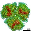 30882MC M: map data used to model this data C: citing same article ( |
|---|---|
| Similar structure data |

-Links
 PDBj
PDBj


- Assembly
| Deposited unit | 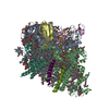
|
|---|---|
| 1 |
|
-Components
-Photosystem I protein ... , 4 types, 4 molecules DFLW
| #1: Protein | Mass: 15114.146 Da / Num. of mol.: 1 / Source method: isolated from a natural source / Source: (natural) Acaryochloris marina MBIC11017 (bacteria) / Strain: MBIC 11017 / References: UniProt: B0C8F1 |
|---|---|
| #6: Protein | Mass: 17844.621 Da / Num. of mol.: 1 / Source method: isolated from a natural source / Source: (natural) Acaryochloris marina MBIC11017 (bacteria) / Strain: MBIC 11017 / References: UniProt: B0C7S7 |
| #8: Protein | Mass: 15349.588 Da / Num. of mol.: 1 / Source method: isolated from a natural source Source: (natural) Acaryochloris marina MBIC 11017 (bacteria)Strain: MBIC 11017 / References: UniProt: B0C7S4 |
| #10: Protein/peptide | Mass: 3648.337 Da / Num. of mol.: 1 / Source method: isolated from a natural source / Source: (natural) Acaryochloris marina MBIC11017 (bacteria) / Strain: MBIC 11017 |
-Photosystem I P740 chlorophyll a apoprotein ... , 2 types, 2 molecules AB
| #2: Protein | Mass: 83470.539 Da / Num. of mol.: 1 / Source method: isolated from a natural source / Source: (natural) Acaryochloris marina MBIC11017 (bacteria) / Strain: MBIC 11017 / References: UniProt: B0C474, photosystem I |
|---|---|
| #3: Protein | Mass: 82210.555 Da / Num. of mol.: 1 / Source method: isolated from a natural source / Source: (natural) Acaryochloris marina MBIC11017 (bacteria) / Strain: MBIC 11017 / References: UniProt: B0C475, photosystem I |
-Protein , 1 types, 1 molecules C
| #4: Protein | Mass: 8825.207 Da / Num. of mol.: 1 / Source method: isolated from a natural source Source: (natural) Acaryochloris marinan MBIC11017 (bacteria)Strain: MBIC 11017 / References: UniProt: B0CB42, photosystem I |
|---|
-Photosystem I reaction center subunit ... , 3 types, 3 molecules EJM
| #5: Protein | Mass: 9560.822 Da / Num. of mol.: 1 / Source method: isolated from a natural source / Source: (natural) Acaryochloris marina MBIC11017 (bacteria) / Strain: MBIC 11017 / References: UniProt: B0C5D5 |
|---|---|
| #7: Protein | Mass: 5900.939 Da / Num. of mol.: 1 / Source method: isolated from a natural source Source: (natural) Acaryochloris marina MBIC 11017 (bacteria)Strain: MBIC 11017 / References: UniProt: B0C7S6 |
| #9: Protein/peptide | Mass: 3190.899 Da / Num. of mol.: 1 / Source method: isolated from a natural source / Source: (natural) Acaryochloris marina MBIC11017 (bacteria) / Strain: MBIC 11017 |
-Non-polymers , 8 types, 100 molecules 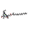


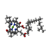

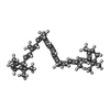

| #11: Chemical | ChemComp-CL7 /  Mass: 895.462 Da / Num. of mol.: 76 / Source method: obtained synthetically / Formula: C54H70MgN4O6 / Feature type: SUBJECT OF INVESTIGATION Mass: 895.462 Da / Num. of mol.: 76 / Source method: obtained synthetically / Formula: C54H70MgN4O6 / Feature type: SUBJECT OF INVESTIGATION#12: Chemical |  Mass: 450.696 Da / Num. of mol.: 2 / Source method: obtained synthetically / Formula: C31H46O2 / Feature type: SUBJECT OF INVESTIGATION Mass: 450.696 Da / Num. of mol.: 2 / Source method: obtained synthetically / Formula: C31H46O2 / Feature type: SUBJECT OF INVESTIGATION#13: Chemical |  Mass: 351.640 Da / Num. of mol.: 3 / Source method: obtained synthetically / Formula: Fe4S4 Mass: 351.640 Da / Num. of mol.: 3 / Source method: obtained synthetically / Formula: Fe4S4#14: Chemical |  Mass: 722.970 Da / Num. of mol.: 2 / Source method: obtained synthetically / Formula: C38H75O10P / Comment: phospholipid*YM Mass: 722.970 Da / Num. of mol.: 2 / Source method: obtained synthetically / Formula: C38H75O10P / Comment: phospholipid*YM#15: Chemical | ChemComp-G9R / |  Mass: 895.462 Da / Num. of mol.: 1 / Source method: obtained synthetically / Formula: C54H70MgN4O6 / Feature type: SUBJECT OF INVESTIGATION Mass: 895.462 Da / Num. of mol.: 1 / Source method: obtained synthetically / Formula: C54H70MgN4O6 / Feature type: SUBJECT OF INVESTIGATION#16: Chemical |  Mass: 871.200 Da / Num. of mol.: 2 / Source method: obtained synthetically / Formula: C55H74N4O5 / Feature type: SUBJECT OF INVESTIGATION Mass: 871.200 Da / Num. of mol.: 2 / Source method: obtained synthetically / Formula: C55H74N4O5 / Feature type: SUBJECT OF INVESTIGATION#17: Chemical | ChemComp-8CT / (  Mass: 536.873 Da / Num. of mol.: 13 / Source method: obtained synthetically / Formula: C40H56 / Feature type: SUBJECT OF INVESTIGATION Mass: 536.873 Da / Num. of mol.: 13 / Source method: obtained synthetically / Formula: C40H56 / Feature type: SUBJECT OF INVESTIGATION#18: Chemical | ChemComp-LMG / |  Mass: 787.158 Da / Num. of mol.: 1 / Source method: obtained synthetically / Formula: C45H86O10 / Feature type: SUBJECT OF INVESTIGATION Mass: 787.158 Da / Num. of mol.: 1 / Source method: obtained synthetically / Formula: C45H86O10 / Feature type: SUBJECT OF INVESTIGATION |
|---|
-Details
| Has ligand of interest | Y |
|---|---|
| Has protein modification | Y |
-Experimental details
-Experiment
| Experiment | Method: ELECTRON MICROSCOPY |
|---|---|
| EM experiment | Aggregation state: PARTICLE / 3D reconstruction method: single particle reconstruction |
- Sample preparation
| Component | Name: Photosystem I from a chlorophyll d-containing cyanobacterium Acaryochloris marina Type: COMPLEX / Entity ID: #1-#10 / Source: NATURAL |
|---|---|
| Source (natural) | Organism: Acaryochloris marina MBIC11017 (bacteria) |
| Buffer solution | pH: 6.5 |
| Specimen | Embedding applied: NO / Shadowing applied: NO / Staining applied: NO / Vitrification applied: YES |
| Specimen support | Grid material: GOLD / Grid mesh size: 300 divisions/in. / Grid type: Quantifoil R2/1 |
| Vitrification | Cryogen name: ETHANE / Humidity: 100 % |
- Electron microscopy imaging
| Experimental equipment |  Model: Titan Krios / Image courtesy: FEI Company |
|---|---|
| Microscopy | Model: FEI TITAN KRIOS |
| Electron gun | Electron source:  FIELD EMISSION GUN / Accelerating voltage: 300 kV / Illumination mode: FLOOD BEAM FIELD EMISSION GUN / Accelerating voltage: 300 kV / Illumination mode: FLOOD BEAM |
| Electron lens | Mode: BRIGHT FIELD / Nominal magnification: 22500 X / Calibrated magnification: 38244 X / Nominal defocus max: 3000 nm / Nominal defocus min: 2500 nm / Calibrated defocus min: 2500 nm / Calibrated defocus max: 3000 nm / Cs: 2.7 mm / C2 aperture diameter: 70 µm / Alignment procedure: COMA FREE |
| Specimen holder | Cryogen: NITROGEN / Specimen holder model: FEI TITAN KRIOS AUTOGRID HOLDER |
| Image recording | Average exposure time: 10 sec. / Electron dose: 47 e/Å2 / Detector mode: SUPER-RESOLUTION / Film or detector model: GATAN K2 SUMMIT (4k x 4k) |
| Image scans | Width: 3710 / Height: 3838 / Movie frames/image: 4 / Used frames/image: 1-40 |
- Processing
| Software | Name: PHENIX / Version: 1.19.1_4122: / Classification: refinement | ||||||||||||||||||||||||
|---|---|---|---|---|---|---|---|---|---|---|---|---|---|---|---|---|---|---|---|---|---|---|---|---|---|
| EM software |
| ||||||||||||||||||||||||
| CTF correction | Type: NONE | ||||||||||||||||||||||||
| Particle selection | Num. of particles selected: 1523356 / Details: raw particles | ||||||||||||||||||||||||
| Symmetry | Point symmetry: C3 (3 fold cyclic) | ||||||||||||||||||||||||
| 3D reconstruction | Resolution: 3.3 Å / Resolution method: FSC 0.143 CUT-OFF / Num. of particles: 240880 / Num. of class averages: 1 / Symmetry type: POINT | ||||||||||||||||||||||||
| Refine LS restraints |
|
