 Movie
Movie Controller

[English] 日本語
 Yorodumi
- PDB-7dvi: Crystal Structure of AbnU: An exo-specific intermolecular Diels-A... -
+ Open data

- Basic information
| Entry | Database: PDB / ID: 7dvi | ||||||||||||
|---|---|---|---|---|---|---|---|---|---|---|---|---|---|
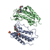
| Title | Crystal Structure of AbnU: An exo-specific intermolecular Diels-Alderase | ||||||||||||
 Components Components | AOC_like domain-containing protein | ||||||||||||
 Keywords Keywords | ISOMERASE / Diels ALderase | ||||||||||||
| Function / homology | Allene oxide cyclase barrel-like domain / Allene oxide cyclase barrel like domain / antibiotic biosynthetic process / isomerase activity / Allene oxide cyclase barrel-like domain-containing protein Function and homology information Function and homology information | ||||||||||||
| Biological species |  Frankia sp. Cc1.17 (bacteria) Frankia sp. Cc1.17 (bacteria) | ||||||||||||
| Method |  X-RAY DIFFRACTION / SYNCHROTRON / MOLECULAR REPLACEMENT / molecular replacement / Resolution: 2 Å X-RAY DIFFRACTION / SYNCHROTRON / MOLECULAR REPLACEMENT / molecular replacement / Resolution: 2 Å | ||||||||||||
 Authors Authors | Kashyap, R. / Addlagatta, A. | ||||||||||||
| Funding support |  India, 3items India, 3items
| ||||||||||||
 Citation Citation | Journal: Commun Chem / Year: 2021 Title: Exo-selective intermolecular Diels-Alder reaction by PyrI4 and AbnU on non-natural substrates. Authors: Kashyap, R. / Yerra, N.V. / Oja, J. / Bala, S. / Potuganti, R. / Thota, J.R. / Alla, M. / Pal, D. / Addlagatta, A. | ||||||||||||
| History |
|
- Structure visualization
| Structure viewer | Molecule: MolmilJmol/JSmol |
|---|
- Downloads & links
-Download
| PDBx/mmCIF format | 7dvi.cif.gz | 47.7 KB | Display | PDBx/mmCIF format |
|---|---|---|---|---|
| PDB format | pdb7dvi.ent.gz | 31.9 KB | Display | PDB format |
| PDBx/mmJSON format | 7dvi.json.gz | Tree view | PDBx/mmJSON format | |
| Others |  Other downloads Other downloads |
-Validation report
| Summary document | 7dvi_validation.pdf.gz | 460.6 KB | Display | wwPDB validaton report |
|---|---|---|---|---|
| Full document | 7dvi_full_validation.pdf.gz | 461.2 KB | Display | |
| Data in XML | 7dvi_validation.xml.gz | 8.9 KB | Display | |
| Data in CIF | 7dvi_validation.cif.gz | 11.5 KB | Display | |
| Arichive directory | https://data.pdbj.org/pub/pdb/validation_reports/dv/7dviftp://data.pdbj.org/pub/pdb/validation_reports/dv/7dvi | HTTPS FTP |
-Related structure data
| Related structure data |  7dvkC  5dyvS S: Starting model for refinement C: citing same article ( |
|---|---|
| Similar structure data |


-Links
 PDBj
PDBj- Assembly
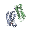
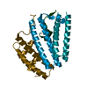
| Deposited unit | 
| ||||||||
|---|---|---|---|---|---|---|---|---|---|
| 1 | 
| ||||||||
| Unit cell |
| ||||||||
| Components on special symmetry positions |
|
-Components
| #1: Protein | Mass: 17071.918 Da / Num. of mol.: 1 Source method: isolated from a genetically manipulated source Source: (gene. exp.) Frankia sp. Cc1.17 (bacteria) / Gene: CC117_14200 / Production host: | ||||||
|---|---|---|---|---|---|---|---|
| #2: Chemical | ChemComp-EDO /   Mass: 62.068 Da / Num. of mol.: 21 / Source method: obtained synthetically / Formula: C2H6O2 Mass: 62.068 Da / Num. of mol.: 21 / Source method: obtained synthetically / Formula: C2H6O2#3: Chemical | ChemComp-DMS / |   Mass: 78.133 Da / Num. of mol.: 1 / Source method: obtained synthetically / Formula: C2H6OS / Comment: DMSO, precipitant*YM Mass: 78.133 Da / Num. of mol.: 1 / Source method: obtained synthetically / Formula: C2H6OS / Comment: DMSO, precipitant*YM#4: Water | ChemComp-HOH / |  Mass: 18.015 Da / Num. of mol.: 69 / Source method: isolated from a natural source / Formula: H2O Mass: 18.015 Da / Num. of mol.: 69 / Source method: isolated from a natural source / Formula: H2OHas ligand of interest | N | |
-Experimental details
-Experiment
| Experiment | Method: X-RAY DIFFRACTION / Number of used crystals: 1 |
|---|
- Sample preparation
| Crystal | Density Matthews: 3.12 Å3/Da / Density % sol: 60.57 % |
|---|---|
| Crystal grow | Temperature: 298 K / Method: vapor diffusion, hanging drop / pH: 7 / Details: 1 M Imidazole, pH 7.0, 20% Ethanol |
-Data collection
| Diffraction | Mean temperature: 100 K / Serial crystal experiment: N | ||||||||||||||||||||||||||||||
|---|---|---|---|---|---|---|---|---|---|---|---|---|---|---|---|---|---|---|---|---|---|---|---|---|---|---|---|---|---|---|---|
| Diffraction source | Source: SYNCHROTRON / Site: ELETTRA  / Beamline: 11.2C / Wavelength: 0.974 Å / Beamline: 11.2C / Wavelength: 0.974 Å | ||||||||||||||||||||||||||||||
| Detector | Type: DECTRIS PILATUS 6M-F / Detector: PIXEL / Date: May 28, 2019 | ||||||||||||||||||||||||||||||
| Radiation | Protocol: SINGLE WAVELENGTH / Monochromatic (M) / Laue (L): M / Scattering type: x-ray | ||||||||||||||||||||||||||||||
| Radiation wavelength | Wavelength: 0.974 Å / Relative weight: 1 | ||||||||||||||||||||||||||||||
| Reflection | Resolution: 2→45.26 Å / Num. obs: 15158 / % possible obs: 99.9 % / Redundancy: 36.4 % / CC1/2: 1 / Rmerge(I) obs: 0.064 / Rpim(I) all: 0.011 / Rrim(I) all: 0.065 / Net I/σ(I): 40.6 | ||||||||||||||||||||||||||||||
| Reflection shell | Diffraction-ID: 1
|
-Phasing
| Phasing | Method: molecular replacement |
|---|
- Processing
| Software |
| ||||||||||||||||||||||||||||||||||||||||||||||||||||||||||||
|---|---|---|---|---|---|---|---|---|---|---|---|---|---|---|---|---|---|---|---|---|---|---|---|---|---|---|---|---|---|---|---|---|---|---|---|---|---|---|---|---|---|---|---|---|---|---|---|---|---|---|---|---|---|---|---|---|---|---|---|---|---|
| Refinement | Method to determine structure: MOLECULAR REPLACEMENT Starting model: 5DYV Resolution: 2→45.22 Å / Cor.coef. Fo:Fc: 0.961 / Cor.coef. Fo:Fc free: 0.942 / SU B: 3.591 / SU ML: 0.097 / Cross valid method: THROUGHOUT / σ(F): 0 / ESU R: 0.143 / ESU R Free: 0.147 / Stereochemistry target values: MAXIMUM LIKELIHOOD Details: HYDROGENS HAVE BEEN ADDED IN THE RIDING POSITIONS U VALUES : REFINED INDIVIDUALLY
| ||||||||||||||||||||||||||||||||||||||||||||||||||||||||||||
| Solvent computation | Ion probe radii: 0.8 Å / Shrinkage radii: 0.8 Å / VDW probe radii: 1.2 Å / Solvent model: MASK | ||||||||||||||||||||||||||||||||||||||||||||||||||||||||||||
| Displacement parameters | Biso max: 91.81 Å2 / Biso mean: 36.25 Å2 / Biso min: 22.22 Å2
| ||||||||||||||||||||||||||||||||||||||||||||||||||||||||||||
| Refinement step | Cycle: final / Resolution: 2→45.22 Å
| ||||||||||||||||||||||||||||||||||||||||||||||||||||||||||||
| Refine LS restraints |
| ||||||||||||||||||||||||||||||||||||||||||||||||||||||||||||
| LS refinement shell | Resolution: 2.003→2.055 Å / Rfactor Rfree error: 0 / Total num. of bins used: 20
|
