 Movie
Movie Controller

[English] 日本語
 Yorodumi
- PDB-1vg7: Crystal Structure Of Octaprenyl Pyrophosphate Synthase From Hyper... -
+ Open data

- Basic information
| Entry | Database: PDB / ID: 1vg7 | ||||||
|---|---|---|---|---|---|---|---|
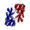
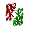
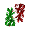
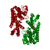
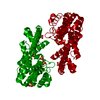
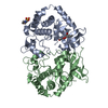
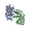
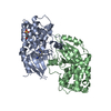
| Title | Crystal Structure Of Octaprenyl Pyrophosphate Synthase From Hyperthermophilic Thermotoga Maritima F132A/L128A/I123A/D62A mutant | ||||||
 Components Components | octoprenyl-diphosphate synthase | ||||||
 Keywords Keywords | TRANSFERASE / trans-type prenyltransferase / thermophilic | ||||||
| Function / homology |  Function and homology information Function and homology informationprenyltransferase activity / isoprenoid biosynthetic process / metal ion binding Similarity search - Function | ||||||
| Biological species |   Thermotoga maritima (bacteria) Thermotoga maritima (bacteria) | ||||||
| Method |  X-RAY DIFFRACTION / SYNCHROTRON / MOLECULAR REPLACEMENT / Resolution: 3.4 Å X-RAY DIFFRACTION / SYNCHROTRON / MOLECULAR REPLACEMENT / Resolution: 3.4 Å | ||||||
 Authors Authors | Guo, R.T. / Kuo, C.J. / Ko, T.P. / Chou, C.C. / Liang, P.H. / Wang, A.H.-J. | ||||||
 Citation Citation | Journal: Biochemistry / Year: 2004 Title: A molecular ruler for chain elongation catalyzed by octaprenyl pyrophosphate synthase and its structure-based engineering to produce unprecedented long chain trans-prenyl products Authors: Guo, R.T. / Kuo, C.J. / Ko, T.P. / Chou, C.C. / Liang, P.H. / Wang, A.H.-J. | ||||||
| History |
|
- Structure visualization
| Structure viewer | Molecule: MolmilJmol/JSmol |
|---|
- Downloads & links
-Download
| PDBx/mmCIF format | 1vg7.cif.gz | 71.8 KB | Display | PDBx/mmCIF format |
|---|---|---|---|---|
| PDB format | pdb1vg7.ent.gz | 53.7 KB | Display | PDB format |
| PDBx/mmJSON format | 1vg7.json.gz | Tree view | PDBx/mmJSON format | |
| Others |  Other downloads Other downloads |
-Validation report
| Arichive directory | https://data.pdbj.org/pub/pdb/validation_reports/vg/1vg7ftp://data.pdbj.org/pub/pdb/validation_reports/vg/1vg7 | HTTPS FTP |
|---|
-Related structure data
| Related structure data |  1vg2C  1vg3C  1vg4C  1vg6C  1v4eS S: Starting model for refinement C: citing same article ( |
|---|---|
| Similar structure data |


-Links
 PDBj
PDBj
- Assembly
| Deposited unit | 
| ||||||||
|---|---|---|---|---|---|---|---|---|---|
| 1 | 
| ||||||||
| 2 | 
| ||||||||
| 3 | x 8
| ||||||||
| Unit cell |
| ||||||||
| Components on special symmetry positions |
|
-Components
| #1: Protein | Mass: 33696.070 Da / Num. of mol.: 1 / Mutation: F132A/L128A/I123A/D62A Source method: isolated from a genetically manipulated source Source: (gene. exp.) Thermotoga maritima (bacteria) / Plasmid: PET32XA-LIC / Species (production host): Escherichia coli / Production host: |
|---|---|
| #2: Water | ChemComp-HOH /  Mass: 18.015 Da / Num. of mol.: 213 / Source method: isolated from a natural source / Formula: H2O Mass: 18.015 Da / Num. of mol.: 213 / Source method: isolated from a natural source / Formula: H2O |
-Experimental details
-Experiment
| Experiment | Method: X-RAY DIFFRACTION / Number of used crystals: 1 |
|---|
- Sample preparation
| Crystal | Density Matthews: 2.86 Å3/Da / Density % sol: 55 % |
|---|---|
| Crystal grow | Temperature: 298 K / Method: vapor diffusion, hanging drop / pH: 7.5 Details: Hepes, Lithium sulfate, PEG 900, pH 7.5, VAPOR DIFFUSION, HANGING DROP, temperature 298.0K |
-Data collection
| Diffraction | Mean temperature: 100 K |
|---|---|
| Diffraction source | Source: SYNCHROTRON / Site: NSRRC  / Beamline: BL17B2 / Wavelength: 1 Å / Beamline: BL17B2 / Wavelength: 1 Å |
| Detector | Type: ADSC QUANTUM 4 / Detector: CCD / Date: Jan 22, 2004 / Details: mirrors |
| Radiation | Monochromator: GRAPHITE / Protocol: SINGLE WAVELENGTH / Monochromatic (M) / Laue (L): M / Scattering type: x-ray |
| Radiation wavelength | Wavelength: 1 Å / Relative weight: 1 |
| Reflection | Resolution: 3.4→30 Å / Num. all: 5586 / Num. obs: 5575 / % possible obs: 99.8 % / Redundancy: 6.47 % / Rmerge(I) obs: 0.12 / Net I/σ(I): 16.78 |
| Reflection shell | Resolution: 3.4→3.52 Å / Rmerge(I) obs: 0.48 / Mean I/σ(I) obs: 3.66 / Num. unique all: 532 / % possible all: 99.8 |
- Processing
| Software |
| |||||||||||||||||||||||||
|---|---|---|---|---|---|---|---|---|---|---|---|---|---|---|---|---|---|---|---|---|---|---|---|---|---|---|
| Refinement | Method to determine structure: MOLECULAR REPLACEMENT Starting model: PDB ENTRY 1V4E Resolution: 3.4→24.77 Å / Isotropic thermal model: Isotropic / Cross valid method: THROUGHOUT / σ(F): 4 / Stereochemistry target values: Engh & Huber
| |||||||||||||||||||||||||
| Displacement parameters | Biso mean: 48.2 Å2 | |||||||||||||||||||||||||
| Refine analyze | Luzzati coordinate error obs: 0.33 Å / Luzzati sigma a obs: 0.32 Å | |||||||||||||||||||||||||
| Refinement step | Cycle: LAST / Resolution: 3.4→24.77 Å
| |||||||||||||||||||||||||
| Refine LS restraints |
| |||||||||||||||||||||||||
| LS refinement shell | Resolution: 3.4→3.52 Å
|
