 Movie
Movie Controller


+ Open data

- Basic information
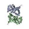
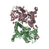
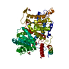
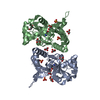
| Entry | Database: PDB / ID: 7dv2 | ||||||
|---|---|---|---|---|---|---|---|
| Title | Structure of Sulfolobus solfataricus SegB-DNA complex | ||||||
 Components Components |
| ||||||
 Keywords Keywords | DNA BINDING PROTEIN / DNA BINDING PROTEIN-DNA COMPLEX | ||||||
| Function / homology | DNA / DNA (> 10) / Uncharacterized protein Function and homology information Function and homology information | ||||||
| Biological species |   Saccharolobus solfataricus (archaea) Saccharolobus solfataricus (archaea)synthetic construct (others) | ||||||
| Method |  X-RAY DIFFRACTION / SYNCHROTRON / MOLECULAR REPLACEMENT / Resolution: 3.1 Å X-RAY DIFFRACTION / SYNCHROTRON / MOLECULAR REPLACEMENT / Resolution: 3.1 Å | ||||||
 Authors Authors | Yen, C.Y. / Lin, M.G. / Sun, Y.J. / Hsiao, C.D. | ||||||
| Funding support |  Taiwan, 1items Taiwan, 1items
| ||||||
 Citation Citation | Journal: Nucleic Acids Res. / Year: 2021 Title: Chromosome segregation in Archaea: SegA- and SegB-DNA complex structures provide insights into segrosome assembly. Authors: Yen, C.Y. / Lin, M.G. / Chen, B.W. / Ng, I.W. / Read, N. / Kabli, A.F. / Wu, C.T. / Shen, Y.Y. / Chen, C.H. / Barilla, D. / Sun, Y.J. / Hsiao, C.D. | ||||||
| History |
|
- Structure visualization
| Structure viewer | Molecule: MolmilJmol/JSmol |
|---|
- Downloads & links
-Download
| PDBx/mmCIF format | 7dv2.cif.gz | 98.2 KB | Display | PDBx/mmCIF format |
|---|---|---|---|---|
| PDB format | pdb7dv2.ent.gz | 71.7 KB | Display | PDB format |
| PDBx/mmJSON format | 7dv2.json.gz | Tree view | PDBx/mmJSON format | |
| Others |  Other downloads Other downloads |
-Validation report
| Arichive directory | https://data.pdbj.org/pub/pdb/validation_reports/dv/7dv2ftp://data.pdbj.org/pub/pdb/validation_reports/dv/7dv2 | HTTPS FTP |
|---|
-Related structure data
| Related structure data |  7dutC 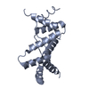 7duvSC  7dv3C 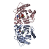 7dwrC S: Starting model for refinement C: citing same article ( |
|---|---|
| Similar structure data |


-Links
 PDBj
PDBj


- Assembly
| Deposited unit | 
| ||||||||
|---|---|---|---|---|---|---|---|---|---|
| 1 |
| ||||||||
| Unit cell |
|
-Components
| #1: Protein | Mass: 9991.653 Da / Num. of mol.: 4 Source method: isolated from a genetically manipulated source Details: N-terminal 33 residues truncated protein Source: (gene. exp.) Saccharolobus solfataricus (strain ATCC 35092 / DSM 1617 / JCM 11322 / P2) (archaea)Strain: ATCC 35092 / DSM 1617 / JCM 11322 / P2 / Gene: SSO0035 / Production host:  #2: DNA chain | | Mass: 6520.249 Da / Num. of mol.: 1 / Source method: obtained synthetically / Details: A21 missing / Source: (synth.) synthetic construct (others) #3: DNA chain | | Mass: 6373.134 Da / Num. of mol.: 1 / Source method: obtained synthetically / Source: (synth.) synthetic construct (others) |
|---|
-Experimental details
-Experiment
| Experiment | Method: X-RAY DIFFRACTION / Number of used crystals: 1 |
|---|
- Sample preparation
| Crystal | Density Matthews: 3.36 Å3/Da / Density % sol: 63.36 % |
|---|---|
| Crystal grow | Temperature: 293 K / Method: vapor diffusion, sitting drop Details: sodium citrate tribasic dihydrate pH 5.0, magnesium chloride hexahydrate, PEG 20000 |
-Data collection
| Diffraction | Mean temperature: 100 K / Serial crystal experiment: N | |||||||||||||||||||||||||||||||||||||||||||||||||||||||||||||||||||||||||||||||||||||||||||||||||||
|---|---|---|---|---|---|---|---|---|---|---|---|---|---|---|---|---|---|---|---|---|---|---|---|---|---|---|---|---|---|---|---|---|---|---|---|---|---|---|---|---|---|---|---|---|---|---|---|---|---|---|---|---|---|---|---|---|---|---|---|---|---|---|---|---|---|---|---|---|---|---|---|---|---|---|---|---|---|---|---|---|---|---|---|---|---|---|---|---|---|---|---|---|---|---|---|---|---|---|---|---|
| Diffraction source | Source: SYNCHROTRON / Site: NSRRC / Beamline: BL15A1 / Wavelength: 1 Å | |||||||||||||||||||||||||||||||||||||||||||||||||||||||||||||||||||||||||||||||||||||||||||||||||||
| Detector | Type: RAYONIX MX300HE / Detector: CCD / Date: Nov 21, 2020 | |||||||||||||||||||||||||||||||||||||||||||||||||||||||||||||||||||||||||||||||||||||||||||||||||||
| Radiation | Protocol: SINGLE WAVELENGTH / Monochromatic (M) / Laue (L): M / Scattering type: x-ray | |||||||||||||||||||||||||||||||||||||||||||||||||||||||||||||||||||||||||||||||||||||||||||||||||||
| Radiation wavelength | Wavelength: 1 Å / Relative weight: 1 | |||||||||||||||||||||||||||||||||||||||||||||||||||||||||||||||||||||||||||||||||||||||||||||||||||
| Reflection | Resolution: 3.1→30 Å / Num. obs: 13274 / % possible obs: 98.3 % / Redundancy: 6.3 % / Rmerge(I) obs: 0.104 / Rpim(I) all: 0.045 / Rrim(I) all: 0.114 / Χ2: 0.851 / Net I/σ(I): 6.8 / Num. measured all: 83127 | |||||||||||||||||||||||||||||||||||||||||||||||||||||||||||||||||||||||||||||||||||||||||||||||||||
| Reflection shell | Diffraction-ID: 1
|
- Processing
| Software |
| ||||||||||||||||||||||||||||||||||||||||||||||||||||||||
|---|---|---|---|---|---|---|---|---|---|---|---|---|---|---|---|---|---|---|---|---|---|---|---|---|---|---|---|---|---|---|---|---|---|---|---|---|---|---|---|---|---|---|---|---|---|---|---|---|---|---|---|---|---|---|---|---|---|
| Refinement | Method to determine structure: MOLECULAR REPLACEMENT Starting model: 7DUV Resolution: 3.1→26.87 Å / SU ML: 0.42 / Cross valid method: THROUGHOUT / σ(F): 1.36 / Phase error: 30.76 / Stereochemistry target values: ML
| ||||||||||||||||||||||||||||||||||||||||||||||||||||||||
| Solvent computation | Shrinkage radii: 0.9 Å / VDW probe radii: 1.11 Å / Solvent model: FLAT BULK SOLVENT MODEL | ||||||||||||||||||||||||||||||||||||||||||||||||||||||||
| Displacement parameters | Biso max: 141.77 Å2 / Biso mean: 55.4415 Å2 / Biso min: 25.34 Å2 | ||||||||||||||||||||||||||||||||||||||||||||||||||||||||
| Refinement step | Cycle: final / Resolution: 3.1→26.87 Å
| ||||||||||||||||||||||||||||||||||||||||||||||||||||||||
| LS refinement shell | Refine-ID: X-RAY DIFFRACTION / Rfactor Rfree error: 0 / Total num. of bins used: 7
|
