 Movie
Movie Controller

[English] 日本語
 Yorodumi
- PDB-7dtn: Crystal structure of metallo-beta-lactamase IMP-1 mutant (D120E) ... -
+ Open data

- Basic information
| Entry | Database: PDB / ID: 7dtn | |||||||||
|---|---|---|---|---|---|---|---|---|---|---|
| Title | Crystal structure of metallo-beta-lactamase IMP-1 mutant (D120E) in complex with citrate. | |||||||||
 Components Components | Metallo-beta-lactamase type 2 | |||||||||
 Keywords Keywords | HYDROLASE / metallo-beta-lactamase / zinc(II) ion / citrate | |||||||||
| Function / homology |  Function and homology information Function and homology informationHydrolases; Acting on ester bonds; Endoribonucleases producing 5'-phosphomonoesters / antibiotic catabolic process / beta-lactamase activity / beta-lactamase / periplasmic space / response to antibiotic / zinc ion binding Similarity search - Function | |||||||||
| Biological species |  Serratia marcescens (bacteria) Serratia marcescens (bacteria) | |||||||||
| Method |  X-RAY DIFFRACTION / SYNCHROTRON / MOLECULAR REPLACEMENT / Resolution: 1.85 Å X-RAY DIFFRACTION / SYNCHROTRON / MOLECULAR REPLACEMENT / Resolution: 1.85 Å | |||||||||
 Authors Authors | Yamaguchi, Y. / Kurosaki, H. | |||||||||
| Funding support |  Japan, 2items Japan, 2items
| |||||||||
 Citation Citation | Journal: J.Med.Chem. / Year: 2021 Title: Crystal Structures of Metallo-beta-Lactamase (IMP-1) and Its D120E Mutant in Complexes with Citrate and the Inhibitory Effect of the Benzyl Group in Citrate Monobenzyl Ester. Authors: Yamaguchi, Y. / Kato, K. / Ichimaru, Y. / Jin, W. / Sakai, M. / Abe, M. / Wachino, J.I. / Arakawa, Y. / Miyagi, Y. / Imai, M. / Fukuishi, N. / Yamagata, Y. / Otsuka, M. / Fujita, M. / Kurosaki, H. | |||||||||
| History |
|
- Structure visualization
| Structure viewer | Molecule: MolmilJmol/JSmol |
|---|
- Downloads & links
-Download
| PDBx/mmCIF format | 7dtn.cif.gz | 198.6 KB | Display | PDBx/mmCIF format |
|---|---|---|---|---|
| PDB format | pdb7dtn.ent.gz | 155.7 KB | Display | PDB format |
| PDBx/mmJSON format | 7dtn.json.gz | Tree view | PDBx/mmJSON format | |
| Others |  Other downloads Other downloads |
-Validation report
| Arichive directory | https://data.pdbj.org/pub/pdb/validation_reports/dt/7dtnftp://data.pdbj.org/pub/pdb/validation_reports/dt/7dtn | HTTPS FTP |
|---|
-Related structure data
| Related structure data |  7dtmC  1dd6S S: Starting model for refinement C: citing same article ( |
|---|---|
| Similar structure data |


-Links
 PDBj
PDBj


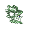
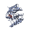
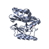
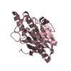
- Assembly
| Deposited unit | 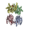
| ||||||||
|---|---|---|---|---|---|---|---|---|---|
| 1 | 
| ||||||||
| 2 | 
| ||||||||
| 3 | 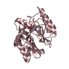
| ||||||||
| 4 | 
| ||||||||
| Unit cell |
|
-Components
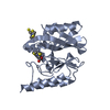
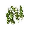
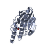
| #1: Protein | Mass: 25208.699 Da / Num. of mol.: 4 / Mutation: D81E Source method: isolated from a genetically manipulated source Source: (gene. exp.) Serratia marcescens (bacteria) / Plasmid: pET9a / Production host: #2: Chemical | ChemComp-ZN /   Mass: 65.409 Da / Num. of mol.: 8 / Source method: obtained synthetically / Formula: Zn / Feature type: SUBJECT OF INVESTIGATION Mass: 65.409 Da / Num. of mol.: 8 / Source method: obtained synthetically / Formula: Zn / Feature type: SUBJECT OF INVESTIGATION#3: Chemical | ChemComp-FLC /   Mass: 189.100 Da / Num. of mol.: 4 / Source method: obtained synthetically / Formula: C6H5O7 / Feature type: SUBJECT OF INVESTIGATION Mass: 189.100 Da / Num. of mol.: 4 / Source method: obtained synthetically / Formula: C6H5O7 / Feature type: SUBJECT OF INVESTIGATION#4: Chemical | ChemComp-AE3 / 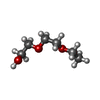  Mass: 134.174 Da / Num. of mol.: 4 / Source method: obtained synthetically / Formula: C6H14O3 Mass: 134.174 Da / Num. of mol.: 4 / Source method: obtained synthetically / Formula: C6H14O3#5: Water | ChemComp-HOH / |  Mass: 18.015 Da / Num. of mol.: 627 / Source method: isolated from a natural source / Formula: H2O Mass: 18.015 Da / Num. of mol.: 627 / Source method: isolated from a natural source / Formula: H2OHas ligand of interest | Y | |
|---|
-Experimental details
-Experiment
| Experiment | Method: X-RAY DIFFRACTION / Number of used crystals: 1 |
|---|
- Sample preparation
| Crystal | Density Matthews: 2.74 Å3/Da / Density % sol: 55.16 % |
|---|---|
| Crystal grow | Temperature: 293 K / Method: vapor diffusion, hanging drop / pH: 7 Details: 0.1M citric acid-sodium citrate buffer, 0.2M sodium acetate, PEG4000 30 w/v% |
-Data collection
| Diffraction | Mean temperature: 100 K / Serial crystal experiment: N |
|---|---|
| Diffraction source | Source: SYNCHROTRON / Site: Photon Factory / Beamline: BL-5A / Wavelength: 1 Å |
| Detector | Type: ADSC QUANTUM 315r / Detector: CCD / Date: Oct 16, 2011 |
| Radiation | Protocol: SINGLE WAVELENGTH / Monochromatic (M) / Laue (L): M / Scattering type: x-ray |
| Radiation wavelength | Wavelength: 1 Å / Relative weight: 1 |
| Reflection | Resolution: 1.85→50 Å / Num. obs: 89488 / % possible obs: 97.7 % / Redundancy: 3.9 % / Rmerge(I) obs: 0.05 / Net I/σ(I): 26.4 |
| Reflection shell | Resolution: 1.85→1.88 Å / Rmerge(I) obs: 0.332 / Mean I/σ(I) obs: 5 / Num. unique obs: 4327 |
- Processing
| Software |
| ||||||||||||||||||||||||||||||||||||||||||||||||||||||||||||
|---|---|---|---|---|---|---|---|---|---|---|---|---|---|---|---|---|---|---|---|---|---|---|---|---|---|---|---|---|---|---|---|---|---|---|---|---|---|---|---|---|---|---|---|---|---|---|---|---|---|---|---|---|---|---|---|---|---|---|---|---|---|
| Refinement | Method to determine structure: MOLECULAR REPLACEMENT Starting model: 1dd6 Resolution: 1.85→30 Å / Cor.coef. Fo:Fc: 0.96 / Cor.coef. Fo:Fc free: 0.943 / SU B: 2.287 / SU ML: 0.07 / Cross valid method: THROUGHOUT / σ(F): 0 / ESU R: 0.116 / ESU R Free: 0.111 / Stereochemistry target values: MAXIMUM LIKELIHOOD Details: HYDROGENS HAVE BEEN ADDED IN THE RIDING POSITIONS U VALUES : REFINED INDIVIDUALLY
| ||||||||||||||||||||||||||||||||||||||||||||||||||||||||||||
| Solvent computation | Ion probe radii: 0.8 Å / Shrinkage radii: 0.8 Å / VDW probe radii: 1.2 Å / Solvent model: MASK | ||||||||||||||||||||||||||||||||||||||||||||||||||||||||||||
| Displacement parameters | Biso max: 70.04 Å2 / Biso mean: 19.461 Å2 / Biso min: 8.68 Å2
| ||||||||||||||||||||||||||||||||||||||||||||||||||||||||||||
| Refinement step | Cycle: final / Resolution: 1.85→30 Å
| ||||||||||||||||||||||||||||||||||||||||||||||||||||||||||||
| Refine LS restraints |
| ||||||||||||||||||||||||||||||||||||||||||||||||||||||||||||
| LS refinement shell | Resolution: 1.85→1.896 Å / Rfactor Rfree error: 0
|
