 Movie
Movie Controller

[English] 日本語
 Yorodumi
- PDB-7dsr: Anthranilate phosphoribosyltransferase variant Gly141Asn from Sac... -
+ Open data

- Basic information
| Entry | Database: PDB / ID: 7dsr | |||||||||
|---|---|---|---|---|---|---|---|---|---|---|
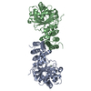
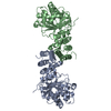
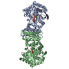
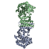
| Title | Anthranilate phosphoribosyltransferase variant Gly141Asn from Saccharomyces cerevisiae in complex with 4-fluoroanthranilate | |||||||||
 Components Components | Anthranilate phosphoribosyltransferase | |||||||||
 Keywords Keywords | TRANSFERASE / Glycosyltransferase / Tryptophan biosynthesis / anthranilate phosphoribosyltransferase activity | |||||||||
| Function / homology |  Function and homology information Function and homology informationanthranilate phosphoribosyltransferase / anthranilate phosphoribosyltransferase activity / L-tryptophan biosynthetic process / nucleus / cytoplasm / cytosol Similarity search - Function | |||||||||
| Biological species |  | |||||||||
| Method |  X-RAY DIFFRACTION / SYNCHROTRON / MOLECULAR REPLACEMENT / Resolution: 2.5 Å X-RAY DIFFRACTION / SYNCHROTRON / MOLECULAR REPLACEMENT / Resolution: 2.5 Å | |||||||||
 Authors Authors | Wu, X. | |||||||||
| Funding support |  China, 2items China, 2items
| |||||||||
 Citation Citation | Journal: Acta Crystallogr.,Sect.F / Year: 2021 Title: Crystal structures of anthranilate phosphoribosyltransferase from Saccharomyces cerevisiae. Authors: Wu, X. / Zhang, M. / Kuang, Z. / Yue, J. / Xue, L. / Zhu, M. / Zhu, Z. / Khan, M.H. / Niu, L. | |||||||||
| History |
|
- Structure visualization
| Structure viewer | Molecule: MolmilJmol/JSmol |
|---|
- Downloads & links
-Download
| PDBx/mmCIF format | 7dsr.cif.gz | 147.8 KB | Display | PDBx/mmCIF format |
|---|---|---|---|---|
| PDB format | pdb7dsr.ent.gz | 113.4 KB | Display | PDB format |
| PDBx/mmJSON format | 7dsr.json.gz | Tree view | PDBx/mmJSON format | |
| Others |  Other downloads Other downloads |
-Validation report
| Arichive directory | https://data.pdbj.org/pub/pdb/validation_reports/ds/7dsrftp://data.pdbj.org/pub/pdb/validation_reports/ds/7dsr | HTTPS FTP |
|---|
-Related structure data
| Related structure data |  7dsjC  7dsmSC  7dsoC  7dspC C: citing same article ( S: Starting model for refinement |
|---|---|
| Similar structure data |

-Links
 PDBj
PDBj- Assembly
| Deposited unit | 
| ||||||||
|---|---|---|---|---|---|---|---|---|---|
| 1 |
| ||||||||
| Unit cell |
|
-Components
| #1: Protein | Mass: 41896.824 Da / Num. of mol.: 2 / Mutation: G141A Source method: isolated from a genetically manipulated source Source: (gene. exp.)  References: UniProt: P07285, anthranilate phosphoribosyltransferase #2: Chemical | ChemComp-FA0 / |   Mass: 155.126 Da / Num. of mol.: 1 / Source method: obtained synthetically / Formula: C7H6FNO2 / Feature type: SUBJECT OF INVESTIGATION Mass: 155.126 Da / Num. of mol.: 1 / Source method: obtained synthetically / Formula: C7H6FNO2 / Feature type: SUBJECT OF INVESTIGATION#3: Water | ChemComp-HOH / |  Mass: 18.015 Da / Num. of mol.: 70 / Source method: isolated from a natural source / Formula: H2O Mass: 18.015 Da / Num. of mol.: 70 / Source method: isolated from a natural source / Formula: H2OHas ligand of interest | Y | |
|---|
-Experimental details
-Experiment
| Experiment | Method: X-RAY DIFFRACTION / Number of used crystals: 1 |
|---|
- Sample preparation
| Crystal | Density Matthews: 2.21 Å3/Da / Density % sol: 44.35 % |
|---|---|
| Crystal grow | Temperature: 287 K / Method: vapor diffusion, sitting drop / Details: 20 % PEG 4000, 0.1 M Tris PH 8.0 |
-Data collection
| Diffraction | Mean temperature: 100 K / Serial crystal experiment: N |
|---|---|
| Diffraction source | Source: SYNCHROTRON / Site: SSRF / Beamline: BL18U1 / Wavelength: 0.97915 Å |
| Detector | Type: DECTRIS PILATUS3 6M / Detector: PIXEL / Date: Jun 20, 2020 |
| Radiation | Protocol: SINGLE WAVELENGTH / Monochromatic (M) / Laue (L): M / Scattering type: x-ray |
| Radiation wavelength | Wavelength: 0.97915 Å / Relative weight: 1 |
| Reflection | Resolution: 2.5→50 Å / Num. obs: 26794 / % possible obs: 99.8 % / Redundancy: 12.9 % / Rpim(I) all: 0.027 / Rrim(I) all: 0.099 / Net I/σ(I): 31.8 |
| Reflection shell | Resolution: 2.5→2.54 Å / Redundancy: 11.9 % / Mean I/σ(I) obs: 4.2 / Num. unique obs: 1311 / Rpim(I) all: 0.145 / Rrim(I) all: 0.507 / % possible all: 100 |
- Processing
| Software |
| ||||||||||||||||||||||||||||||||||||||||||||||||||||||||||||
|---|---|---|---|---|---|---|---|---|---|---|---|---|---|---|---|---|---|---|---|---|---|---|---|---|---|---|---|---|---|---|---|---|---|---|---|---|---|---|---|---|---|---|---|---|---|---|---|---|---|---|---|---|---|---|---|---|---|---|---|---|---|
| Refinement | Method to determine structure: MOLECULAR REPLACEMENT Starting model: 7DSM Resolution: 2.5→46.36 Å / Cor.coef. Fo:Fc: 0.932 / Cor.coef. Fo:Fc free: 0.902 / SU B: 11.129 / SU ML: 0.248 / Cross valid method: THROUGHOUT / σ(F): 0 / ESU R: 0.639 / ESU R Free: 0.315 / Stereochemistry target values: MAXIMUM LIKELIHOOD Details: HYDROGENS HAVE BEEN ADDED IN THE RIDING POSITIONS U VALUES : REFINED INDIVIDUALLY
| ||||||||||||||||||||||||||||||||||||||||||||||||||||||||||||
| Solvent computation | Ion probe radii: 0.8 Å / Shrinkage radii: 0.8 Å / VDW probe radii: 1.2 Å / Solvent model: MASK | ||||||||||||||||||||||||||||||||||||||||||||||||||||||||||||
| Displacement parameters | Biso max: 108.76 Å2 / Biso mean: 46.108 Å2 / Biso min: 25.8 Å2
| ||||||||||||||||||||||||||||||||||||||||||||||||||||||||||||
| Refinement step | Cycle: final / Resolution: 2.5→46.36 Å
| ||||||||||||||||||||||||||||||||||||||||||||||||||||||||||||
| Refine LS restraints |
| ||||||||||||||||||||||||||||||||||||||||||||||||||||||||||||
| LS refinement shell | Resolution: 2.5→2.565 Å / Rfactor Rfree error: 0 / Total num. of bins used: 20
|
