 Movie
Movie Controller

[English] 日本語
 Yorodumi
- PDB-7dsj: Anthranilate phosphoribosyltransferase from Saccharomyces cerevis... -
+ Open data

- Basic information
| Entry | Database: PDB / ID: 7dsj | |||||||||
|---|---|---|---|---|---|---|---|---|---|---|
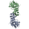
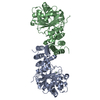
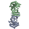
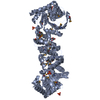
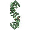
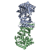
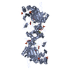
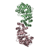
| Title | Anthranilate phosphoribosyltransferase from Saccharomyces cerevisiae in complex with PRPP and Mg | |||||||||
 Components Components | Anthranilate phosphoribosyltransferase | |||||||||
 Keywords Keywords | TRANSFERASE / Glycosyltransferase / Tryptophan biosynthesis | |||||||||
| Function / homology |  Function and homology information Function and homology informationanthranilate phosphoribosyltransferase / anthranilate phosphoribosyltransferase activity / L-tryptophan biosynthetic process / nucleus / cytoplasm / cytosol Similarity search - Function | |||||||||
| Biological species |  | |||||||||
| Method |  X-RAY DIFFRACTION / SYNCHROTRON / MOLECULAR REPLACEMENT / Resolution: 2.44 Å X-RAY DIFFRACTION / SYNCHROTRON / MOLECULAR REPLACEMENT / Resolution: 2.44 Å | |||||||||
 Authors Authors | Wu, X. | |||||||||
| Funding support |  China, 2items China, 2items
| |||||||||
 Citation Citation | Journal: Acta Crystallogr.,Sect.F / Year: 2021 Title: Crystal structures of anthranilate phosphoribosyltransferase from Saccharomyces cerevisiae. Authors: Wu, X. / Zhang, M. / Kuang, Z. / Yue, J. / Xue, L. / Zhu, M. / Zhu, Z. / Khan, M.H. / Niu, L. | |||||||||
| History |
|
- Structure visualization
| Structure viewer | Molecule: MolmilJmol/JSmol |
|---|
- Downloads & links
-Download
| PDBx/mmCIF format | 7dsj.cif.gz | 153.4 KB | Display | PDBx/mmCIF format |
|---|---|---|---|---|
| PDB format | pdb7dsj.ent.gz | 118.6 KB | Display | PDB format |
| PDBx/mmJSON format | 7dsj.json.gz | Tree view | PDBx/mmJSON format | |
| Others |  Other downloads Other downloads |
-Validation report
| Arichive directory | https://data.pdbj.org/pub/pdb/validation_reports/ds/7dsjftp://data.pdbj.org/pub/pdb/validation_reports/ds/7dsj | HTTPS FTP |
|---|
-Related structure data
| Related structure data |  7dsmSC  7dsoC  7dspC  7dsrC S: Starting model for refinement C: citing same article ( |
|---|---|
| Similar structure data |

-Links
 PDBj
PDBj
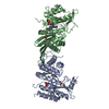
- Assembly
| Deposited unit | 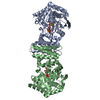
| ||||||||
|---|---|---|---|---|---|---|---|---|---|
| 1 |
| ||||||||
| Unit cell |
|
-Components
| #1: Protein | Mass: 41701.625 Da / Num. of mol.: 2 Source method: isolated from a genetically manipulated source Source: (gene. exp.)  References: UniProt: P07285, anthranilate phosphoribosyltransferase #2: Sugar |   Type: D-saccharide / Mass: 390.070 Da / Num. of mol.: 2 / Source method: obtained synthetically / Formula: C5H13O14P3 / Feature type: SUBJECT OF INVESTIGATION Type: D-saccharide / Mass: 390.070 Da / Num. of mol.: 2 / Source method: obtained synthetically / Formula: C5H13O14P3 / Feature type: SUBJECT OF INVESTIGATION#3: Chemical |   Mass: 24.305 Da / Num. of mol.: 2 / Source method: obtained synthetically / Formula: Mg / Feature type: SUBJECT OF INVESTIGATION Mass: 24.305 Da / Num. of mol.: 2 / Source method: obtained synthetically / Formula: Mg / Feature type: SUBJECT OF INVESTIGATION#4: Water | ChemComp-HOH / |  Mass: 18.015 Da / Num. of mol.: 133 / Source method: isolated from a natural source / Formula: H2O Mass: 18.015 Da / Num. of mol.: 133 / Source method: isolated from a natural source / Formula: H2OHas ligand of interest | Y | |
|---|
-Experimental details
-Experiment
| Experiment | Method: X-RAY DIFFRACTION / Number of used crystals: 1 |
|---|
- Sample preparation
| Crystal | Density Matthews: 2.26 Å3/Da / Density % sol: 45.62 % |
|---|---|
| Crystal grow | Temperature: 287 K / Method: vapor diffusion, sitting drop / pH: 6.5 / Details: 0.1 M BIS-TRIS PH 6.5, 16% W/V PEG 10000 |
-Data collection
| Diffraction | Mean temperature: 100 K / Serial crystal experiment: N |
|---|---|
| Diffraction source | Source: SYNCHROTRON / Site: SSRF / Beamline: BL18U1 / Wavelength: 0.9793 Å |
| Detector | Type: DECTRIS PILATUS3 6M / Detector: PIXEL / Date: Dec 28, 2019 |
| Radiation | Protocol: SINGLE WAVELENGTH / Monochromatic (M) / Laue (L): M / Scattering type: x-ray |
| Radiation wavelength | Wavelength: 0.9793 Å / Relative weight: 1 |
| Reflection | Resolution: 2.44→50 Å / Num. obs: 29444 / % possible obs: 100 % / Redundancy: 13.1 % / Rpim(I) all: 0.027 / Rrim(I) all: 0.097 / Net I/σ(I): 24.3 |
| Reflection shell | Resolution: 2.45→2.49 Å / Redundancy: 12.9 % / Mean I/σ(I) obs: 4 / Num. unique obs: 1440 / Rpim(I) all: 0.134 / Rrim(I) all: 0.484 / % possible all: 100 |
- Processing
| Software |
| ||||||||||||||||||||||||||||||||||||||||||||||||||||||||||||
|---|---|---|---|---|---|---|---|---|---|---|---|---|---|---|---|---|---|---|---|---|---|---|---|---|---|---|---|---|---|---|---|---|---|---|---|---|---|---|---|---|---|---|---|---|---|---|---|---|---|---|---|---|---|---|---|---|---|---|---|---|---|
| Refinement | Method to determine structure: MOLECULAR REPLACEMENT Starting model: 7DSM Resolution: 2.44→46.99 Å / Cor.coef. Fo:Fc: 0.958 / Cor.coef. Fo:Fc free: 0.936 / SU B: 8.965 / SU ML: 0.204 / Cross valid method: THROUGHOUT / σ(F): 0 / ESU R: 0.468 / ESU R Free: 0.271 / Stereochemistry target values: MAXIMUM LIKELIHOOD Details: HYDROGENS HAVE BEEN ADDED IN THE RIDING POSITIONS U VALUES : REFINED INDIVIDUALLY
| ||||||||||||||||||||||||||||||||||||||||||||||||||||||||||||
| Solvent computation | Ion probe radii: 0.8 Å / Shrinkage radii: 0.8 Å / VDW probe radii: 1.2 Å / Solvent model: MASK | ||||||||||||||||||||||||||||||||||||||||||||||||||||||||||||
| Displacement parameters | Biso max: 102.56 Å2 / Biso mean: 41.681 Å2 / Biso min: 23.53 Å2
| ||||||||||||||||||||||||||||||||||||||||||||||||||||||||||||
| Refinement step | Cycle: final / Resolution: 2.44→46.99 Å
| ||||||||||||||||||||||||||||||||||||||||||||||||||||||||||||
| Refine LS restraints |
| ||||||||||||||||||||||||||||||||||||||||||||||||||||||||||||
| LS refinement shell | Resolution: 2.44→2.502 Å / Rfactor Rfree error: 0
|
