 Movie
Movie Controller

[English] 日本語
 Yorodumi
- PDB-7dn2: Acidic stable capsid structure of Helicobacter pylori bacteriopha... -
+ Open data

- Basic information
| Entry | Database: PDB / ID: 7dn2 | ||||||||||||||||||||||||||||||||||||
|---|---|---|---|---|---|---|---|---|---|---|---|---|---|---|---|---|---|---|---|---|---|---|---|---|---|---|---|---|---|---|---|---|---|---|---|---|---|
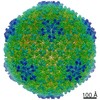
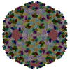
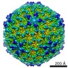
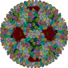
| Title | Acidic stable capsid structure of Helicobacter pylori bacteriophage KHP30 | ||||||||||||||||||||||||||||||||||||
 Components Components |
| ||||||||||||||||||||||||||||||||||||
 Keywords Keywords | VIRUS / CAPSID / PHAGE / PHAGE HEAD / CRYOEM | ||||||||||||||||||||||||||||||||||||
| Function / homology | Protein of unknown function DUF4043 / Phage capsid protein / Major structural protein ORF14 / Uncharacterized protein Function and homology information Function and homology information | ||||||||||||||||||||||||||||||||||||
| Biological species |  Helicobacter pylori bacteriophage KHP30 (virus) Helicobacter pylori bacteriophage KHP30 (virus) | ||||||||||||||||||||||||||||||||||||
| Method | ELECTRON MICROSCOPY / single particle reconstruction / cryo EM / Resolution: 2.7 Å | ||||||||||||||||||||||||||||||||||||
 Authors Authors | Kamiya, R. / Uchiyama, J. / Matsuzaki, S. / Murata, K. / Iwasaki, K. / Miyazaki, N. | ||||||||||||||||||||||||||||||||||||
| Funding support |  Japan, 2items Japan, 2items
| ||||||||||||||||||||||||||||||||||||
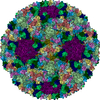
 Citation Citation | Journal: Structure / Year: 2022 Title: Acid-stable capsid structure of Helicobacter pylori bacteriophage KHP30 by single-particle cryoelectron microscopy. Authors: Ryosuke Kamiya / Jumpei Uchiyama / Shigenobu Matsuzaki / Kazuyoshi Murata / Kenji Iwasaki / Naoyuki Miyazaki / Abstract: The acid-stable capsid structures of Helicobacter pylori phages KHP30 and KHP40 are solved at 2.7 and 3.0 Å resolutions by cryoelectron microscopy, respectively. The capsids have icosahedral T = 9 ...The acid-stable capsid structures of Helicobacter pylori phages KHP30 and KHP40 are solved at 2.7 and 3.0 Å resolutions by cryoelectron microscopy, respectively. The capsids have icosahedral T = 9 symmetry and consist of each 540 copies of 2 structural proteins, a major capsid protein, and a cement protein. The major capsid proteins form 12 pentagonal capsomeres occupying icosahedral vertexes and 80 hexagonal capsomeres located at icosahedral faces and edges. The major capsid protein has a unique protruding loop extending to the neighboring subunit that stabilizes hexagonal capsomeres. Furthermore, the capsid is decorated with trimeric cement proteins with a jelly roll motif. The cement protein trimer sits on the quasi-three-fold axis formed by three major capsid protein capsomeres, thereby enhancing the particle stability by connecting these capsomeres. Sequence and structure comparisons between the related Helicobacter pylori phages suggest a possible mechanism of phage adaptation to the human gastric environment. | ||||||||||||||||||||||||||||||||||||
| History |
|
- Structure visualization
| Movie |
Movie viewer |
|---|---|
| Structure viewer | Molecule: MolmilJmol/JSmol |
- Downloads & links
-Download
| PDBx/mmCIF format | 7dn2.cif.gz | 733.9 KB | Display | PDBx/mmCIF format |
|---|---|---|---|---|
| PDB format | pdb7dn2.ent.gz | 618.7 KB | Display | PDB format |
| PDBx/mmJSON format | 7dn2.json.gz | Tree view | PDBx/mmJSON format | |
| Others |  Other downloads Other downloads |
-Validation report
| Arichive directory | https://data.pdbj.org/pub/pdb/validation_reports/dn/7dn2ftp://data.pdbj.org/pub/pdb/validation_reports/dn/7dn2 | HTTPS FTP |
|---|
-Related structure data
| Related structure data | 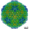 30778MC 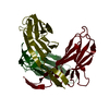 7douC 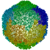 7f2pC M: map data used to model this data C: citing same article ( |
|---|---|
| Similar structure data |
-Links
 PDBj
PDBj- Assembly
| Deposited unit | 
|
|---|---|
| 1 | x 60
|
| 2 |
|
| 3 | x 5
|
| 4 | x 6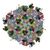
|
| 5 | 
|
| Symmetry | Point symmetry: (Schoenflies symbol: I (icosahedral)) |
-Components
| #1: Protein | Mass: 42465.922 Da / Num. of mol.: 9 / Source method: isolated from a natural source Source: (natural) Helicobacter pylori bacteriophage KHP30 (virus)References: UniProt: I7H0H9 #2: Protein | Mass: 13721.461 Da / Num. of mol.: 9 / Source method: isolated from a natural source Source: (natural) Helicobacter pylori bacteriophage KHP30 (virus)References: UniProt: I7HFW5 Has protein modification | N | |
|---|
-Experimental details
-Experiment
| Experiment | Method: ELECTRON MICROSCOPY |
|---|---|
| EM experiment | Aggregation state: PARTICLE / 3D reconstruction method: single particle reconstruction |
- Sample preparation
| Component | Name: Helicobacter phage KHP / Type: VIRUS / Entity ID: all / Source: NATURAL |
|---|---|
| Molecular weight | Experimental value: NO |
| Source (natural) | Organism: Helicobacter phage KHP (virus) |
| Details of virus | Empty: NO / Enveloped: NO / Isolate: OTHER / Type: VIRION |
| Virus shell | Name: Head / Diameter: 700 nm / Triangulation number (T number): 9 |
| Buffer solution | pH: 7.2 |
| Specimen | Embedding applied: NO / Shadowing applied: NO / Staining applied: NO / Vitrification applied: YES |
| Vitrification | Instrument: FEI VITROBOT MARK IV / Cryogen name: ETHANE / Humidity: 100 % / Chamber temperature: 277 K |
- Electron microscopy imaging
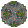
| Experimental equipment |  Model: Titan Krios / Image courtesy: FEI Company |
|---|---|
| Microscopy | Model: FEI TITAN KRIOS |
| Electron gun | Electron source:  FIELD EMISSION GUN / Accelerating voltage: 300 kV / Illumination mode: FLOOD BEAM FIELD EMISSION GUN / Accelerating voltage: 300 kV / Illumination mode: FLOOD BEAM |
| Electron lens | Mode: BRIGHT FIELD |
| Image recording | Average exposure time: 1 sec. / Electron dose: 20 e/Å2 / Film or detector model: FEI FALCON II (4k x 4k) |
- Processing
| Software | Name: PHENIX / Version: 1.16_3549: / Classification: refinement | ||||||||||||||||||||||||
|---|---|---|---|---|---|---|---|---|---|---|---|---|---|---|---|---|---|---|---|---|---|---|---|---|---|
| EM software |
| ||||||||||||||||||||||||
| CTF correction | Type: PHASE FLIPPING AND AMPLITUDE CORRECTION | ||||||||||||||||||||||||
| Symmetry | Point symmetry: I (icosahedral) | ||||||||||||||||||||||||
| 3D reconstruction | Resolution: 2.7 Å / Resolution method: FSC 0.143 CUT-OFF / Num. of particles: 20737 / Algorithm: FOURIER SPACE / Symmetry type: POINT | ||||||||||||||||||||||||
| Refine LS restraints |
|
