Movie
Movie Controller

[English] 日本語
 Yorodumi
- PDB-2cse: Features of Reovirus Outer-Capsid Protein mu1 Revealed by Electro... -
+ Open data

- Basic information
| Entry | Database: PDB / ID: 2cse | ||||||
|---|---|---|---|---|---|---|---|
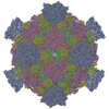
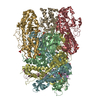
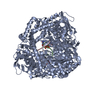
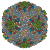
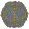
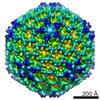
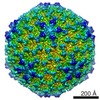
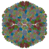
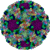
| Title | Features of Reovirus Outer-Capsid Protein mu1 Revealed by Electron and Image Reconstruction of the virion at 7.0-A Resolution | ||||||
 Components Components |
| ||||||
 Keywords Keywords | VIRUS / cryoEM / image processing / reovirus / membrane penetration protein / Icosahedral virus | ||||||
| Function / homology |  Function and homology information Function and homology informationicosahedral viral capsid / T=2 icosahedral viral capsid / host cell surface binding / viral inner capsid / symbiont-mediated suppression of host PKR/eIFalpha signaling / viral outer capsid / symbiont entry into host cell via permeabilization of host membrane / host cell endoplasmic reticulum / host cell mitochondrion / 7-methylguanosine mRNA capping ...icosahedral viral capsid / T=2 icosahedral viral capsid / host cell surface binding / viral inner capsid / symbiont-mediated suppression of host PKR/eIFalpha signaling / viral outer capsid / symbiont entry into host cell via permeabilization of host membrane / host cell endoplasmic reticulum / host cell mitochondrion / 7-methylguanosine mRNA capping / protein serine/threonine kinase inhibitor activity / viral life cycle / viral genome replication / viral capsid / viral nucleocapsid / mRNA guanylyltransferase / mRNA guanylyltransferase activity / mRNA (guanine-N7)-methyltransferase / mRNA 5'-cap (guanine-N7-)-methyltransferase activity / RNA helicase activity / symbiont-mediated suppression of host innate immune response / RNA helicase / symbiont-mediated suppression of host type I interferon-mediated signaling pathway / RNA-directed RNA polymerase / nucleotide binding / hydrolase activity / RNA-directed RNA polymerase activity / GTP binding / host cell plasma membrane / structural molecule activity / ATP hydrolysis activity / RNA binding / zinc ion binding / ATP binding Similarity search - Function | ||||||
| Biological species |  Mammalian orthoreovirus 1Mammalian orthoreovirus 3 Mammalian orthoreovirus 1Mammalian orthoreovirus 3 | ||||||
| Method | ELECTRON MICROSCOPY / single particle reconstruction / cryo EM / Resolution: 7 Å | ||||||
 Authors Authors | Zhang, X. / Ji, Y. / Zhang, L. / Harrison, S.C. / Marinescu, D.C. / Nibert, M.L. / Baker, T.S. | ||||||
 Citation Citation | Journal: Structure / Year: 2005 Title: Features of reovirus outer capsid protein mu1 revealed by electron cryomicroscopy and image reconstruction of the virion at 7.0 Angstrom resolution. Authors: Xing Zhang / Yongchang Ji / Lan Zhang / Stephen C Harrison / Dan C Marinescu / Max L Nibert / Timothy S Baker /  Abstract: Reovirus is a useful model for addressing the molecular basis of membrane penetration by one of the larger nonenveloped animal viruses. We now report the structure of the reovirus virion at ...Reovirus is a useful model for addressing the molecular basis of membrane penetration by one of the larger nonenveloped animal viruses. We now report the structure of the reovirus virion at approximately 7.0 A resolution as obtained by electron cryomicroscopy and three-dimensional image reconstruction. Several features of the myristoylated outer capsid protein mu1, not seen in a previous X-ray crystal structure of the mu1-sigma3 heterohexamer, are evident in the virion. These features appear to be important for stabilizing the outer capsid, regulating the conformational changes in mu1 that accompany perforation of target membranes, and contributing directly to membrane penetration during cell entry. | ||||||
| History |
|
- Structure visualization
| Movie |
Movie viewer |
|---|---|
| Structure viewer | Molecule: MolmilJmol/JSmol |
- Downloads & links
-Download
| PDBx/mmCIF format | 2cse.cif.gz | 519.8 KB | Display | PDBx/mmCIF format |
|---|---|---|---|---|
| PDB format | pdb2cse.ent.gz | 345.4 KB | Display | PDB format |
| PDBx/mmJSON format | 2cse.json.gz | Tree view | PDBx/mmJSON format | |
| Others |  Other downloads Other downloads |
-Validation report
| Arichive directory | https://data.pdbj.org/pub/pdb/validation_reports/cs/2cseftp://data.pdbj.org/pub/pdb/validation_reports/cs/2cse | HTTPS FTP |
|---|
-Related structure data
| Similar structure data |
|---|
-Links
 PDBj
PDBj


- Assembly
| Deposited unit | 
|
|---|---|
| 1 | x 60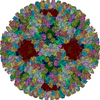
|
| 2 |
|
| 3 | x 5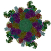
|
| 4 | x 6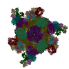
|
| 5 | 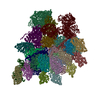
|
| Symmetry | Point symmetry: (Hermann–Mauguin notation: 532 / Schoenflies symbol: I (icosahedral)) |
-Components
-Protein , 6 types, 27 molecules ABCPQRJKLTSDEFMNOGHIUVWXYZ1
| #1: Protein | Mass: 76346.336 Da / Num. of mol.: 10 / Source method: isolated from a natural source / Source: (natural) Mammalian orthoreovirus 1 / Genus: Orthoreovirus / Species: Mammalian orthoreovirus / Strain: Lang / References: UniProt: P11077#2: Protein | Mass: 41237.117 Da / Num. of mol.: 10 / Source method: isolated from a natural source / Source: (natural) Mammalian orthoreovirus 1 / Genus: Orthoreovirus / Species: Mammalian orthoreovirus / Strain: Lang / References: UniProt: P07939#3: Protein | | Mass: 144098.766 Da / Num. of mol.: 1 / Source method: isolated from a natural source / Source: (natural) Mammalian orthoreovirus 3 / Genus: Orthoreovirus / Species: Mammalian orthoreovirus / Strain: Dearing / References: UniProt: P11079#4: Protein | Mass: 142008.359 Da / Num. of mol.: 2 / Source method: isolated from a natural source / Source: (natural) Mammalian orthoreovirus 1 / Genus: Orthoreovirus / Species: Mammalian orthoreovirus / Strain: Lang / References: UniProt: Q9WAB2#5: Protein | Mass: 47155.211 Da / Num. of mol.: 3 / Source method: isolated from a natural source / Source: (natural) Mammalian orthoreovirus 1 / Genus: Orthoreovirus / Species: Mammalian orthoreovirus / Strain: Lang / References: UniProt: P11314#6: Protein | | Mass: 142510.062 Da / Num. of mol.: 1 / Source method: isolated from a natural source / Source: (natural) Mammalian orthoreovirus 1 / Genus: Orthoreovirus / Species: Mammalian orthoreovirus / Strain: Lang / References: UniProt: P17376, UniProt: P0CK32*PLUS |
|---|
-Experimental details
-Experiment
| Experiment | Method: ELECTRON MICROSCOPY |
|---|---|
| EM experiment | Aggregation state: PARTICLE / 3D reconstruction method: single particle reconstruction |
- Sample preparation
| Component | Name: HUMAN REOVIRUS VIRIONS (T3D) / Type: VIRUS / Details: The structure was monodisperse. |
|---|---|
| Details of virus | Host category: MAMMALIAN |
| Buffer solution | Name: 10 mM TRIS / pH: 7.5 / Details: 10 mM TRIS |
| Specimen | Conc.: 2 mg/ml / Embedding applied: NO / Shadowing applied: NO / Staining applied: NO / Vitrification applied: YES |
| Specimen support | Details: HOLEY CARBON |
| Vitrification | Cryogen name: ETHANE / Details: PLUNGED INTO ETHANE |
- Electron microscopy imaging
| EM imaging | Electron source:
| |||||||||||||||||||||||||||||||||||||||
|---|---|---|---|---|---|---|---|---|---|---|---|---|---|---|---|---|---|---|---|---|---|---|---|---|---|---|---|---|---|---|---|---|---|---|---|---|---|---|---|---|
| Image recording | Electron dose: 20 e/Å2 / Film or detector model: KODAK SO-163 FILM | |||||||||||||||||||||||||||||||||||||||
| Image scans | Num. digital images: 54 |
 FIELD EMISSION GUN
- Processing
| CTF correction | Details: CTF correction of each particle | ||||||||||||||||||||||||||||
|---|---|---|---|---|---|---|---|---|---|---|---|---|---|---|---|---|---|---|---|---|---|---|---|---|---|---|---|---|---|
| Symmetry | Point symmetry: I (icosahedral) | ||||||||||||||||||||||||||||
| 3D reconstruction | Method: PFT, OOR, POR / Resolution: 7 Å / Num. of particles: 7939 / Nominal pixel size: 2.3 Å / Actual pixel size: 2.21 Å / Magnification calibration: CROSS-CORRELATION / Symmetry type: POINT | ||||||||||||||||||||||||||||
| Atomic model building | Space: REAL | ||||||||||||||||||||||||||||
| Atomic model building |
| ||||||||||||||||||||||||||||
| Refinement step | Cycle: LAST
|