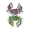
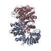
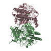
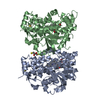
| Deposited unit | A: 3,5-diaminohexanoate dehydrogenase
B: 3,5-diaminohexanoate dehydrogenase
C: 3,5-diaminohexanoate dehydrogenase
D: 3,5-diaminohexanoate dehydrogenase
hetero molecules
| Theoretical mass | Number of molelcules |
|---|
| Total (without water) | 157,951 | 6 |
|---|
| Polymers | 156,464 | 4 |
|---|
| Non-polymers | 1,487 | 2 |
|---|
| Water | 3,387 | 188 |
|---|
|
|---|
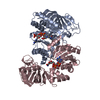
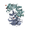
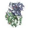
| 1 | A: 3,5-diaminohexanoate dehydrogenase
C: 3,5-diaminohexanoate dehydrogenase
hetero molecules
| Theoretical mass | Number of molelcules |
|---|
| Total (without water) | 79,719 | 4 |
|---|
| Polymers | 78,232 | 2 |
|---|
| Non-polymers | 1,487 | 2 |
|---|
| Water | 36 | 2 |
|---|
| Type | Name | Symmetry operation | Number |
|---|
| identity operation | 1_555 | x,y,z | 1 |
| Buried area | 6340 Å2 |
|---|
| ΔGint | -35 kcal/mol |
|---|
| Surface area | 27730 Å2 |
|---|
| Method | PISA |
|---|
|
|---|
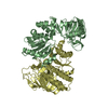
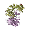
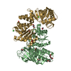
| 2 | B: 3,5-diaminohexanoate dehydrogenase
D: 3,5-diaminohexanoate dehydrogenase
| Theoretical mass | Number of molelcules |
|---|
| Total (without water) | 78,232 | 2 |
|---|
| Polymers | 78,232 | 2 |
|---|
| Non-polymers | 0 | 0 |
|---|
| Water | 36 | 2 |
|---|
| Type | Name | Symmetry operation | Number |
|---|
| identity operation | 1_555 | x,y,z | 1 |
| Buried area | 3860 Å2 |
|---|
| ΔGint | -32 kcal/mol |
|---|
| Surface area | 28560 Å2 |
|---|
| Method | PISA |
|---|
|
|---|
| Unit cell | | Length a, b, c (Å) | 106.241, 127.874, 116.994 |
|---|
| Angle α, β, γ (deg.) | 90.000, 96.295, 90.000 |
|---|
| Int Tables number | 5 |
|---|
| Space group name H-M | C121 |
|---|
| Space group name Hall | C2y |
|---|
| Symmetry operation | #1: x,y,z
#2: -x,y,-z
#3: x+1/2,y+1/2,z
#4: -x+1/2,y+1/2,-z |
|---|
|
|---|
| Noncrystallographic symmetry (NCS) | NCS domain:
NCS domain segments: Ens-ID: 1 | Dom-ID | Component-ID | Beg auth comp-ID | Beg label comp-ID | End auth comp-ID | End label comp-ID | Selection details | Auth asym-ID | Label asym-ID | Auth seq-ID | Label seq-ID |
|---|
| 1 | 1 | METMETHISHIS(chain 'A' and (resid 1 through 11 or resid 13 through 351))AA| 1 - 11 | 1 - 11 | | 1 | 2 | VALVALTYRTYR(chain 'A' and (resid 1 through 11 or resid 13 through 351))AA| 13 - 350 | 13 - 350 | | 2 | 3 | METMETHISHIS(chain 'B' and (resid 1 through 11 or resid 13 through 351))BB| 1 - 11 | 1 - 11 | | 2 | 4 | VALVALTYRTYR(chain 'B' and (resid 1 through 11 or resid 13 through 351))BB| 13 - 350 | 13 - 350 | | 3 | 5 | METMETHISHIS(chain 'C' and (resid 1 through 11 or resid 13 through 351))CC| 1 - 11 | 1 - 11 | | 3 | 6 | VALVALTYRTYR| (chain 'C' and (resid 1 through 11 | | | | | | | | | | | | | | | | | | | | | | | | | | | | | | | | | | | | | | | |
|
|---|
 Movie
Movie Controller

 Yorodumi
 Open data
 Basic information
 Components
 Keywords
 Function and homology information
 X-RAY DIFFRACTION /
  Authors
 China, 1items
  Citation
 Structure visualization
 Downloads & links
 Other downloads
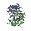


 PDBj
PDBj

 Assembly