 Movie
Movie Controller


+ Open data

- Basic information
| Entry | Database: PDB / ID: 7dgo | ||||||||||||
|---|---|---|---|---|---|---|---|---|---|---|---|---|---|
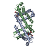
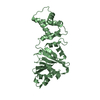
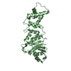
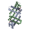
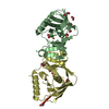
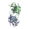
| Title | The Zn-bound dimeric structure of K79H/G80A/H81A myoglobin | ||||||||||||
 Components Components | Myoglobin | ||||||||||||
 Keywords Keywords | OXYGEN BINDING / OXYGEN STORAGE | ||||||||||||
| Function / homology |  Function and homology information Function and homology informationOxidoreductases; Acting on other nitrogenous compounds as donors / nitrite reductase activity / oxygen transport / sarcoplasm / Oxidoreductases; Acting on a peroxide as acceptor; Peroxidases / skeletal muscle contraction / removal of superoxide radicals / oxygen carrier activity / peroxidase activity / oxygen binding ...Oxidoreductases; Acting on other nitrogenous compounds as donors / nitrite reductase activity / oxygen transport / sarcoplasm / Oxidoreductases; Acting on a peroxide as acceptor; Peroxidases / skeletal muscle contraction / removal of superoxide radicals / oxygen carrier activity / peroxidase activity / oxygen binding / heme binding / metal ion binding Similarity search - Function | ||||||||||||
| Biological species |  | ||||||||||||
| Method |  X-RAY DIFFRACTION / SYNCHROTRON / MOLECULAR REPLACEMENT / Resolution: 2 Å X-RAY DIFFRACTION / SYNCHROTRON / MOLECULAR REPLACEMENT / Resolution: 2 Å | ||||||||||||
 Authors Authors | Nagao, S. / Idomoto, A. / Shibata, N. / Higuchi, Y. / Hirota, S. | ||||||||||||
| Funding support |  Japan, 3items Japan, 3items
| ||||||||||||
 Citation Citation | Journal: J.Inorg.Biochem. / Year: 2021 Title: Rational design of metal-binding sites in domain-swapped myoglobin dimers. Authors: Nagao, S. / Idomoto, A. / Shibata, N. / Higuchi, Y. / Hirota, S. | ||||||||||||
| History |
|
- Structure visualization
| Structure viewer | Molecule: MolmilJmol/JSmol |
|---|
- Downloads & links
-Download
| PDBx/mmCIF format | 7dgo.cif.gz | 79.7 KB | Display | PDBx/mmCIF format |
|---|---|---|---|---|
| PDB format | pdb7dgo.ent.gz | 58.2 KB | Display | PDB format |
| PDBx/mmJSON format | 7dgo.json.gz | Tree view | PDBx/mmJSON format | |
| Others |  Other downloads Other downloads |
-Validation report
| Arichive directory | https://data.pdbj.org/pub/pdb/validation_reports/dg/7dgoftp://data.pdbj.org/pub/pdb/validation_reports/dg/7dgo | HTTPS FTP |
|---|
-Related structure data
| Related structure data | 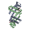 7dgjC  7dgkC 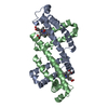 7dglC 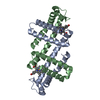 7dgmC  7dgnC 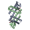 3vm9S S: Starting model for refinement C: citing same article ( |
|---|---|
| Similar structure data |


-Links
 PDBj
PDBj


- Assembly
| Deposited unit | 
| ||||||||||||||||||
|---|---|---|---|---|---|---|---|---|---|---|---|---|---|---|---|---|---|---|---|
| 1 |
| ||||||||||||||||||
| Unit cell |
| ||||||||||||||||||
| Noncrystallographic symmetry (NCS) | NCS domain:
NCS domain segments: Component-ID: _ / Ens-ID: 1 / Beg auth comp-ID: GLY / Beg label comp-ID: GLY / End auth comp-ID: GLY / End label comp-ID: GLY / Refine code: _ / Auth seq-ID: 1 - 153 / Label seq-ID: 1 - 153
|
-Components
| #1: Protein | Mass: 16939.438 Da / Num. of mol.: 2 / Mutation: K79H, G80A, H81A Source method: isolated from a genetically manipulated source Source: (gene. exp.)  #2: Chemical |   Mass: 616.487 Da / Num. of mol.: 2 / Source method: obtained synthetically / Formula: C34H32FeN4O4 / Feature type: SUBJECT OF INVESTIGATION Mass: 616.487 Da / Num. of mol.: 2 / Source method: obtained synthetically / Formula: C34H32FeN4O4 / Feature type: SUBJECT OF INVESTIGATION#3: Chemical |   Mass: 15.999 Da / Num. of mol.: 2 / Source method: obtained synthetically / Formula: O Mass: 15.999 Da / Num. of mol.: 2 / Source method: obtained synthetically / Formula: O#4: Chemical | ChemComp-ZN /   Mass: 65.409 Da / Num. of mol.: 5 / Source method: obtained synthetically / Formula: Zn / Feature type: SUBJECT OF INVESTIGATION Mass: 65.409 Da / Num. of mol.: 5 / Source method: obtained synthetically / Formula: Zn / Feature type: SUBJECT OF INVESTIGATION#5: Water | ChemComp-HOH / |  Mass: 18.015 Da / Num. of mol.: 94 / Source method: isolated from a natural source / Formula: H2O Mass: 18.015 Da / Num. of mol.: 94 / Source method: isolated from a natural source / Formula: H2OHas ligand of interest | Y | |
|---|
-Experimental details
-Experiment
| Experiment | Method: X-RAY DIFFRACTION / Number of used crystals: 1 |
|---|
- Sample preparation
| Crystal | Density Matthews: 1.95 Å3/Da / Density % sol: 37.01 % |
|---|---|
| Crystal grow | Temperature: 277 K / Method: vapor diffusion, sitting drop / pH: 7 Details: 0.1 M Sodium acetate, 0.1 M Tris-HCl, 25% (w/v) PEG 6,000 |
-Data collection
| Diffraction | Mean temperature: 100 K / Serial crystal experiment: N |
|---|---|
| Diffraction source | Source: SYNCHROTRON / Site: SPring-8 / Beamline: BL38B1 / Wavelength: 1 Å |
| Detector | Type: DECTRIS PILATUS3 S 6M / Detector: PIXEL / Date: Dec 2, 2018 |
| Radiation | Protocol: SINGLE WAVELENGTH / Monochromatic (M) / Laue (L): M / Scattering type: x-ray |
| Radiation wavelength | Wavelength: 1 Å / Relative weight: 1 |
| Reflection | Resolution: 2→43.98 Å / Num. obs: 18568 / % possible obs: 100 % / Redundancy: 6.3 % / CC1/2: 0.999 / Rmerge(I) obs: 0.068 / Net I/σ(I): 17.7 |
| Reflection shell | Resolution: 2→2.05 Å / Redundancy: 6.6 % / Rmerge(I) obs: 0.732 / Mean I/σ(I) obs: 2.3 / Num. unique obs: 1354 / CC1/2: 0.772 / % possible all: 99.9 |
- Processing
| Software |
| ||||||||||||||||||||||||||||||||||||||||||||||||||||||||||||||||||||||||||||||||||||||||||||||||||||||||||||||||||||||||||||||||||||||||||||||||||||||||||||||||||||||||||||||||||||||
|---|---|---|---|---|---|---|---|---|---|---|---|---|---|---|---|---|---|---|---|---|---|---|---|---|---|---|---|---|---|---|---|---|---|---|---|---|---|---|---|---|---|---|---|---|---|---|---|---|---|---|---|---|---|---|---|---|---|---|---|---|---|---|---|---|---|---|---|---|---|---|---|---|---|---|---|---|---|---|---|---|---|---|---|---|---|---|---|---|---|---|---|---|---|---|---|---|---|---|---|---|---|---|---|---|---|---|---|---|---|---|---|---|---|---|---|---|---|---|---|---|---|---|---|---|---|---|---|---|---|---|---|---|---|---|---|---|---|---|---|---|---|---|---|---|---|---|---|---|---|---|---|---|---|---|---|---|---|---|---|---|---|---|---|---|---|---|---|---|---|---|---|---|---|---|---|---|---|---|---|---|---|---|---|
| Refinement | Method to determine structure: MOLECULAR REPLACEMENT Starting model: 3VM9 Resolution: 2→43.98 Å / Cor.coef. Fo:Fc: 0.947 / Cor.coef. Fo:Fc free: 0.926 / SU B: 5.539 / SU ML: 0.153 / Cross valid method: THROUGHOUT / ESU R: 0.263 / ESU R Free: 0.208 / Stereochemistry target values: MAXIMUM LIKELIHOOD / Details: HYDROGENS HAVE BEEN ADDED IN THE RIDING POSITIONS
| ||||||||||||||||||||||||||||||||||||||||||||||||||||||||||||||||||||||||||||||||||||||||||||||||||||||||||||||||||||||||||||||||||||||||||||||||||||||||||||||||||||||||||||||||||||||
| Solvent computation | Ion probe radii: 0.8 Å / Shrinkage radii: 0.8 Å / VDW probe radii: 1.2 Å / Solvent model: MASK | ||||||||||||||||||||||||||||||||||||||||||||||||||||||||||||||||||||||||||||||||||||||||||||||||||||||||||||||||||||||||||||||||||||||||||||||||||||||||||||||||||||||||||||||||||||||
| Displacement parameters | Biso mean: 33.233 Å2
| ||||||||||||||||||||||||||||||||||||||||||||||||||||||||||||||||||||||||||||||||||||||||||||||||||||||||||||||||||||||||||||||||||||||||||||||||||||||||||||||||||||||||||||||||||||||
| Refinement step | Cycle: 1 / Resolution: 2→43.98 Å
| ||||||||||||||||||||||||||||||||||||||||||||||||||||||||||||||||||||||||||||||||||||||||||||||||||||||||||||||||||||||||||||||||||||||||||||||||||||||||||||||||||||||||||||||||||||||
| Refine LS restraints |
|